Deposition Date
1991-06-24
Release Date
1992-07-15
Last Version Date
2024-02-14
Entry Detail
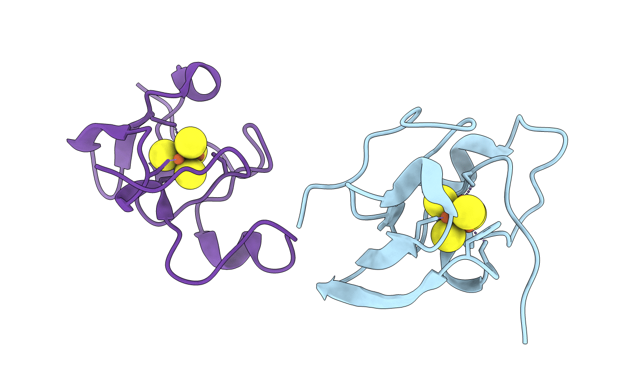
PDB ID:
2HIP
Keywords:
Title:
THE MOLECULAR STRUCTURE OF THE HIGH POTENTIAL IRON-SULFUR PROTEIN ISOLATED FROM ECTOTHIORHODOSPIRA HALOPHILA DETERMINED AT 2.5-ANGSTROMS RESOLUTION
Biological Source:
Source Organism(s):
Halorhodospira halophila (Taxon ID: 1053)
Method Details:
Experimental Method:
Resolution:
2.50 Å
R-Value Observed:
0.18
Space Group:
P 1 21 1