2HIH image
Deposition Date 2006-06-29
Release Date 2007-05-22
Last Version Date 2023-10-25
Entry Detail
PDB ID:
2HIH
Keywords:
Title:
Crystal structure of Staphylococcus hyicus lipase
Biological Source:
Source Organism:
Method Details:
Experimental Method:
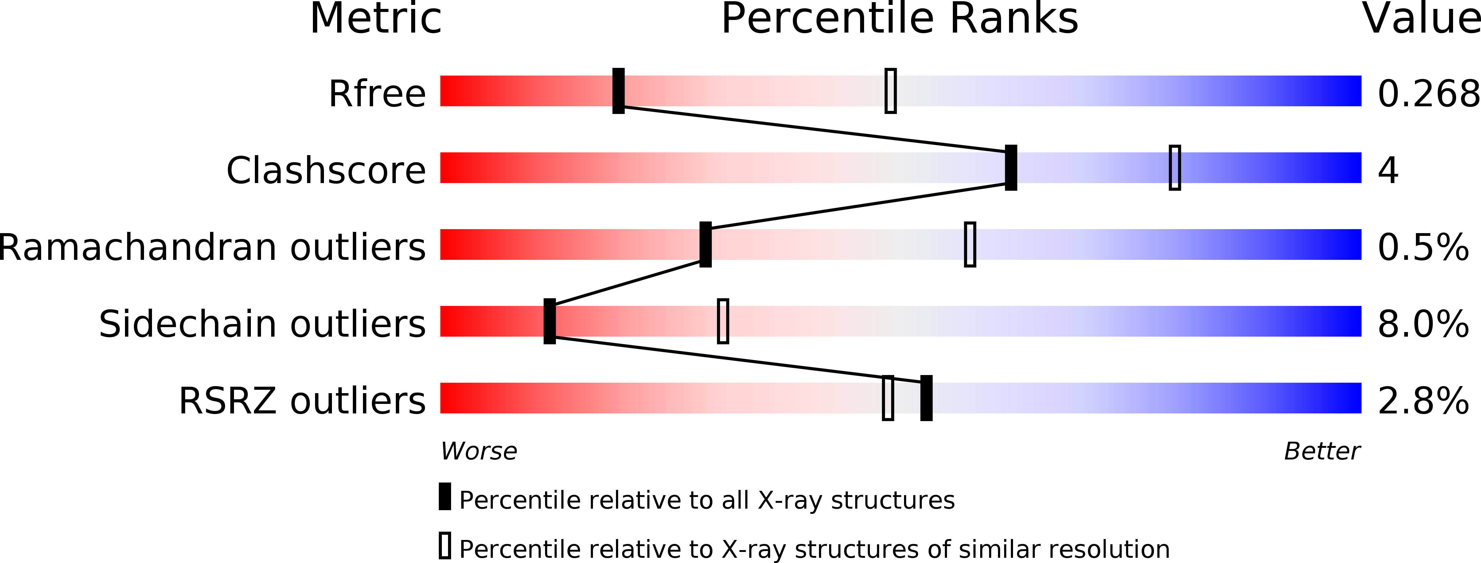
Resolution:
2.86 Å
R-Value Free:
0.26
R-Value Work:
0.20
R-Value Observed:
0.21
Space Group:
P 21 21 21
Macromolecular Entities
Structures with similar UniProt ID
Protein Blast
Polymer Type:polypeptide(L)
Molecule:Lipase 46 kDa form
Gene (Uniprot):lip
Chain IDs:A, B
Chain Length:431
Number of Molecules:2
Biological Source:Staphylococcus hyicus
Primary Citation
Structural basis of phospholipase activity of Staphylococcus hyicus lipase.
J.Mol.Biol. 371 447 456 (2007)
PMID: 17582438 DOI: 10.1016/j.jmb.2007.05.041

Abstact

Staphylococcus hyicus lipase differs from other bacterial lipases in its high phospholipase A1 activity. Here, we present the crystal structure of the S. hyicus lipase at 2.86 A resolution. The lipase is in an open conformation, with the active site partly covered by a neighbouring molecule. Ser124, Asp314 and His355 form the catalytic triad. The substrate-binding cavity contains two large hydrophobic acyl chain-binding pockets and a shallow and more polar third pocket that is capable of binding either a (short) fatty acid or a phospholipid head-group. A model of a phospholipid bound in the active site shows that Lys295 is at hydrogen bonding distance from the substrate's phosphate group. Residues Ser356, Glu292 and Thr294 hold the lysine in position by hydrogen bonding and electrostatic interactions. These observations explain the biochemical data showing the importance of Lys295 and Ser356 for phospholipid binding and phospholipase A1 activity.

Legend

Protein

Chemical

Disease

Primary Citation of related structures
Feedback Form
Name
Email
Institute
Feedback