
Deposition Date
2006-06-28
Release Date
2006-10-24
Last Version Date
2025-03-26
Entry Detail
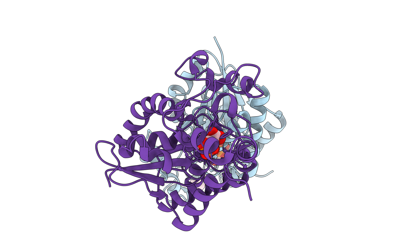
PDB ID:
2HHJ
Keywords:
Title:
Human bisphosphoglycerate mutase complexed with 2,3-bisphosphoglycerate (15 days)
Biological Source:
Source Organism(s):
Homo sapiens (Taxon ID: 9606)
Expression System(s):
Method Details:
Experimental Method:
Resolution:
1.50 Å
R-Value Free:
0.20
R-Value Work:
0.18
R-Value Observed:
0.18
Space Group:
P 21 21 21
