2HH7 image
Deposition Date 2006-06-27
Release Date 2006-12-05
Last Version Date 2024-02-14
Entry Detail
PDB ID:
2HH7
Title:
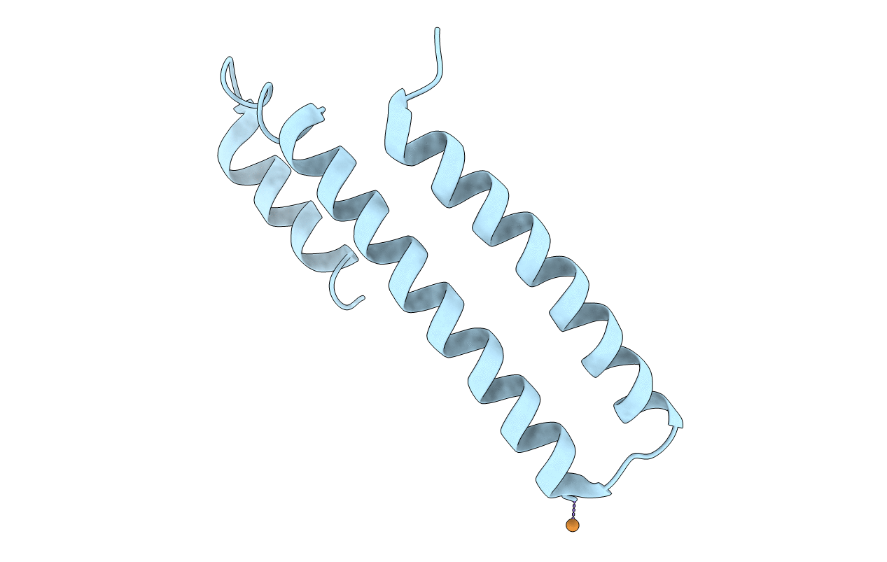
Crystal Structure of Cu(I) bound CsoR from Mycobacterium tuberculosis.
Biological Source:
Source Organism:
Host Organism:
Method Details:
Experimental Method:
Resolution:
2.55 Å
R-Value Free:
0.27
R-Value Work:
0.23
Space Group:
P 64 2 2
Macromolecular Entities
Structures with similar UniProt ID
Protein Blast
Polymer Type:polypeptide(L)
Molecule:Hypothetical protein CsoR
Chain IDs:A
Chain Length:119
Number of Molecules:1
Biological Source:Mycobacterium tuberculosis
Ligand Molecules
Primary Citation
CsoR is a novel Mycobacterium tuberculosis copper-sensing transcriptional regulator.
Nat.Chem.Biol. 3 60 68 (2007)
PMID: 17143269 DOI: 10.1038/nchembio844

Abstact

Copper is an essential element that becomes highly cytotoxic when concentrations exceed the capacity of cells to sequester the ion. Here, we identify a new copper-specific repressor (CsoR) of a copper-sensitive operon (cso) in Mycobacterium tuberculosis (Mtb) that is representative of a large, previously uncharacterized family of proteins (DUF156). Electronic and X-ray absorption spectroscopies reveal that CsoR binds a single-monomer mole equivalent of Cu(I) to form a trigonally coordinated (S(2)N) Cu(I) complex. The 2.6-A crystal structure of copper-loaded CsoR shows a homodimeric antiparallel four-helix bundle architecture that represents a novel DNA-binding fold. The Cu(I) is coordinated by Cys36, Cys65' and His61' in a subunit bridging site. Cu(I) binding negatively regulates the binding of CsoR to a DNA fragment encompassing the operator-promoter region of the Mtb cso operon; this results in derepression of the operon in Mtb and the heterologous host Mycobacterium smegmatis. Substitution of Cys36 or His61 with alanine abolishes Cu(I)- and CsoR-dependent regulation in vivo and in vitro. Potential roles of CsoR in Mtb pathogenesis are discussed.

Legend

Protein

Chemical

Disease

Primary Citation of related structures