2HFT image
Deposition Date 1995-09-08
Release Date 1996-01-29
Last Version Date 2024-11-06
Entry Detail
PDB ID:
2HFT
Title:
THE CRYSTAL STRUCTURE OF THE EXTRACELLULAR DOMAIN OF HUMAN TISSUE FACTOR AT 1.7 ANGSTROMS RESOLUTION
Biological Source:
Source Organism:
Homo sapiens (Taxon ID: 9606)
Host Organism:
Method Details:
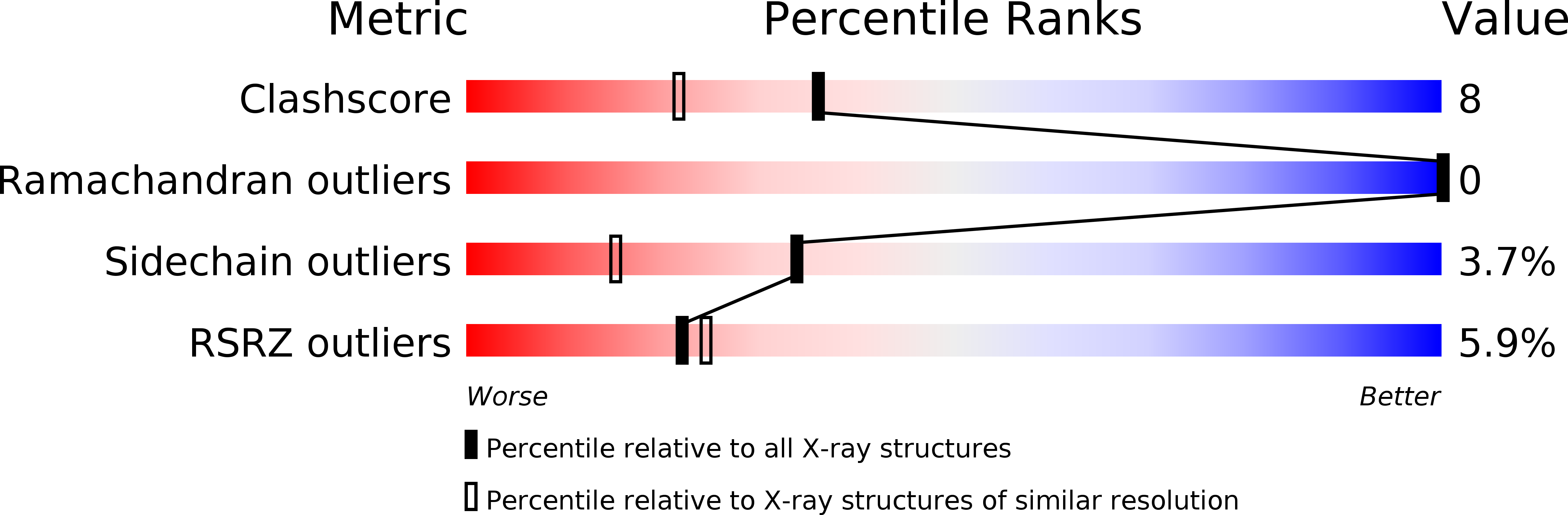
Experimental Method:
Resolution:
1.69 Å
R-Value Free:
0.25
R-Value Observed:
0.20
Space Group:
P 41 21 2
Macromolecular Entities
Polymer Type:polypeptide(L)
Molecule:HUMAN TISSUE FACTOR
Gene (Uniprot):F3
Chain IDs:A
Chain Length:218
Number of Molecules:1
Biological Source:Homo sapiens
Ligand Molecules
Primary Citation
The crystal structure of the extracellular domain of human tissue factor refined to 1.7 A resolution.
J.Mol.Biol. 256 144 159 (1996)
PMID: 8609606 DOI: 10.1006/jmbi.1996.0073

Abstact

Exposure of blood to cells expressing tissue factor results in formation of a high-affinity complex with factor VIIa, initiating the extrinsic pathway of blood coagulation by the activation of factors IX and X. The structure of the extracellular portion of tissue factor was refined to a crystallographic R-value of 20.4% to a resolution of 1.69 A against synchroton data collected from a flash-frozen crystal. The structure consists of two fibronectin type III modules whose hydrophobic cores merge in the domain-domain interface, suggesting that the extracellular portion serves as a relatively rigid template for factor VIIa binding. Analysis of the hydrophobic core of each individual module identifies a cluster of residues forming a packing motif centered on Trp25 which appears to be characteristic for fibronectin type III modules. Comparison of the structure to that of the human growth hormone receptor, which belongs to a different class (class I) of the same cytokine receptor superfamily, shows that the structure of the individual domains is very similar but that the relative domain-domain orientation differs greatly. Even though the WSXWS box characteristic of the class I cytokine receptors is not present in tissue factor, the analogous residues have the identical polyproline helical conformation. Mapping of residues important for biological activity on the structure shows that all these are located on Beta-strands in a small number of distinct clusters, on the opposite side of the molecule compared to the ligand binding determinants of the growth hormone receptor.

Legend

Protein

Chemical

Disease

Primary Citation of related structures