2HF9 image
Deposition Date 2006-06-23
Release Date 2006-07-04
Last Version Date 2024-10-16
Entry Detail
PDB ID:
2HF9
Title:
Crystal structure of HypB from Methanocaldococcus jannaschii in the triphosphate form
Biological Source:
Source Organism:
Host Organism:
Method Details:
Experimental Method:
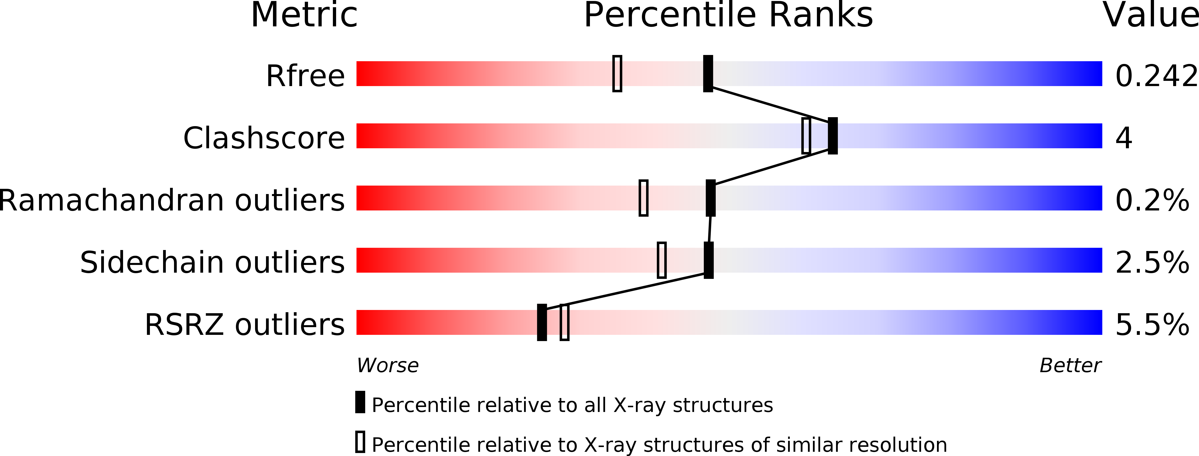
Resolution:
1.90 Å
R-Value Free:
0.24
R-Value Work:
0.21
Space Group:
P 21 21 21
Macromolecular Entities
Polymer Type:polypeptide(L)
Molecule:Probable hydrogenase nickel incorporation protein hypB
Gene (Uniprot):hypB
Chain IDs:A, B
Chain Length:226
Number of Molecules:2
Biological Source:Methanocaldococcus jannaschii
Primary Citation
Structural insights into HypB, a GTP-binding protein that regulates metal binding.
J.Biol.Chem. 281 27492 27502 (2006)
PMID: 16807243 DOI: 10.1074/jbc.M600809200

Abstact

HypB is a prokaryotic metal-binding guanine nucleotide-binding protein that is essential for nickel incorporation into hydrogenases. Here we solved the x-ray structure of HypB from Methanocaldococcus jannaschii. It shows that the G-domain has a different topology than the Ras-like proteins and belongs to the SIMIBI (after Signal Recognition Particle, MinD and BioD) class of NTP-binding proteins. We show that HypB undergoes nucleotide-dependent dimerization, which is apparently a common feature of SIMIBI class G-proteins. The nucleotides are located in the dimer interface and are contacted by both subunits. The active site features residues from both subunits arguing that hydrolysis also requires dimerization. Two metal-binding sites are found, one of which is dependent on the state of bound nucleotide. A totally conserved ENV/IGNLV/ICP motif in switch II relays the nucleotide binding with the metal ionbinding site. The homology with NifH, the Fe protein subunit of nitrogenase, suggests a mechanistic model for the switch-dependent incorporation of a metal ion into hydrogenases.

Legend

Protein

Chemical

Disease

Primary Citation of related structures