2HEZ image
Entry Detail
PDB ID:
2HEZ
Keywords:
Title:
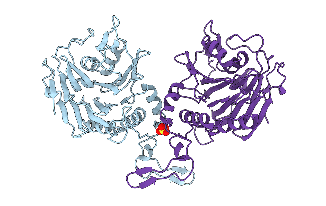
Bifidobacterium longum bile salt hydrolase
Biological Source:
Source Organism:
PDB Version:
Deposition Date:
2006-06-22
Release Date:
2006-09-19
Method Details:
Experimental Method:
Resolution:
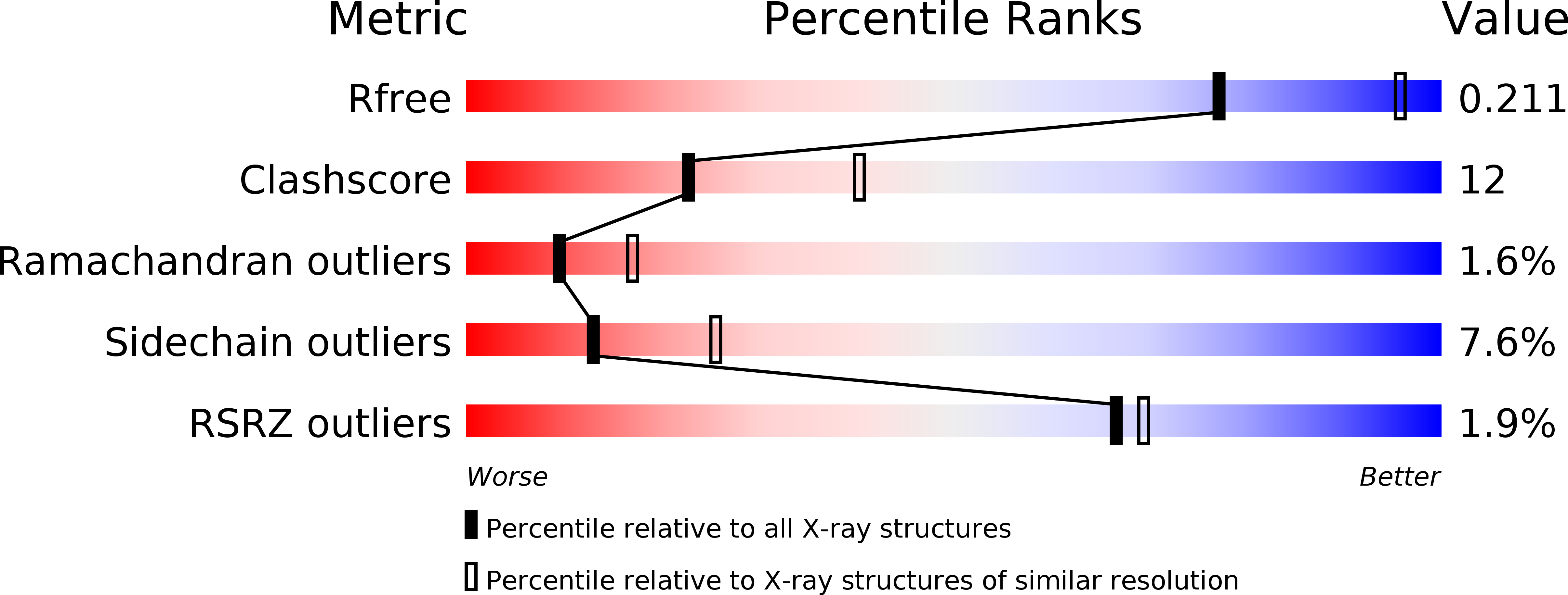
2.50 Å
R-Value Free:
0.21
R-Value Work:
0.17
R-Value Observed:
0.17
Space Group:
P 32 2 1
Macromolecular Entities
Polymer Type:polypeptide(L)
Description:Bile salt hydrolase
Chain IDs:A, B
Chain Length:316
Number of Molecules:2
Biological Source:Bifidobacterium longum
Modified Residue
Compound ID Chain ID Parent Comp ID Details 2D Image
OCS A CYS CYSTEINESULFONIC ACID
Ligand Molecules
Primary Citation
Structural and Functional Analysis of a Conjugated Bile Salt Hydrolase from Bifidobacterium longum Reveals an Evolutionary Relationship with Penicillin V Acylase.
J.Biol.Chem. 281 32516 32525 (2006)
PMID: 16905539 DOI: 10.1074/jbc.M604172200

Abstact

Bile salt hydrolase (BSH) is an enzyme produced by the intestinal microflora that catalyzes the deconjugation of glycine- or taurine-linked bile salts. The crystal structure of BSH reported here from Bifidobacterium longum reveals that it is a member of N-terminal nucleophil hydrolase structural superfamily possessing the characteristic alphabetabetaalpha tetra-lamellar tertiary structure arrangement. Site-directed mutagenesis of the catalytic nucleophil residue, however, shows that it has no role in zymogen processing into its corresponding active form. Substrate specificity was studied using Michaelis-Menten and inhibition kinetics and fluorescence spectroscopy. These data were compared with the specificity profile of BSH from Clostridium perfrigens and pencillin V acylase from Bacillus sphaericus, for both of which the three-dimensional structures are available. Comparative analysis shows a gradation in activity toward common substrates, throwing light on a possible common route toward the evolution of pencillin V acylase and BSH.

Legend

Protein

Chemical

Disease

Primary Citation of related structures