
Deposition Date
2006-06-21
Release Date
2007-02-13
Last Version Date
2024-02-14
Entry Detail
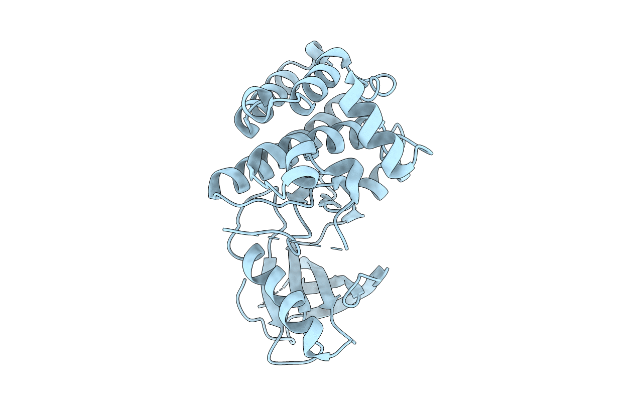
PDB ID:
2HEL
Keywords:
Title:
Crystal structure of a mutant EphA4 kinase domain (Y742A)
Biological Source:
Source Organism(s):
Mus musculus (Taxon ID: 10090)
Expression System(s):
Method Details:
Experimental Method:
Resolution:
2.35 Å
R-Value Free:
0.24
R-Value Work:
0.20
Space Group:
P 1 21 1
