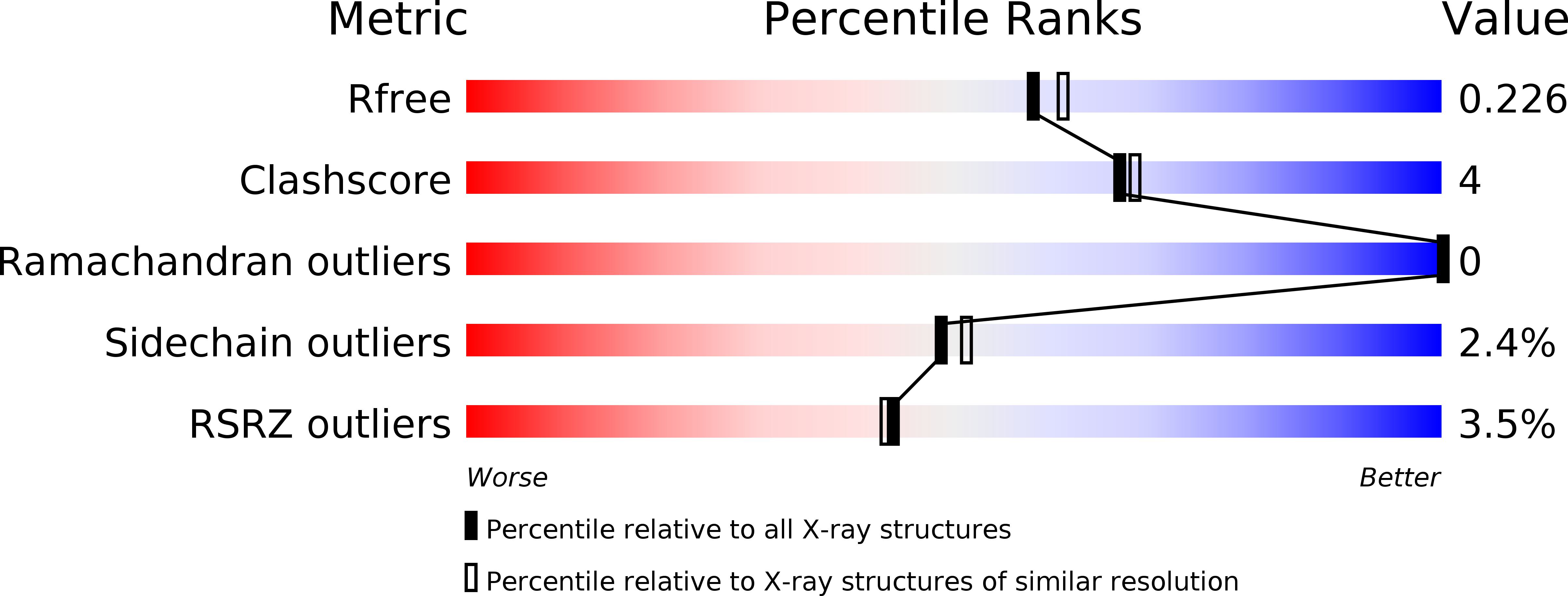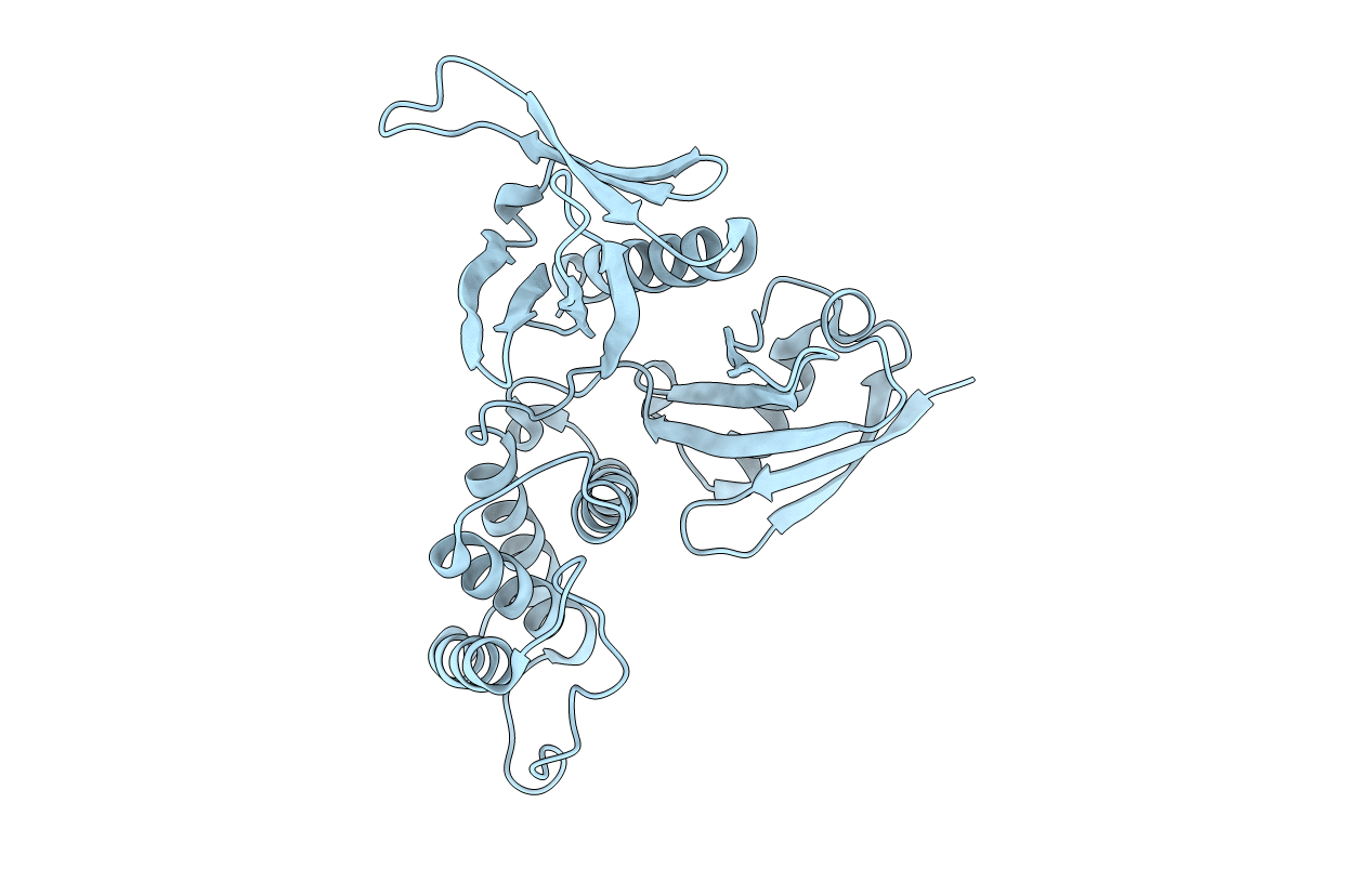
2HE7 image
Deposition Date 2006-06-21
Release Date 2006-07-04
Last Version Date 2024-02-14
Entry Detail
PDB ID:
2HE7
Keywords:
Title:
FERM domain of EPB41L3 (DAL-1)
Biological Source:
Source Organism:
Homo sapiens (Taxon ID: 9606)
Host Organism:
Method Details:
Experimental Method:
Resolution:
2.00 Å
R-Value Free:
0.21
R-Value Work:
0.18
R-Value Observed:
0.18
Space Group:
P 65
Macromolecular Entities
Structures with similar UniProt ID
Protein Blast
Polymer Type:polypeptide(L)
Molecule:Band 4.1-like protein 3
Gene (Uniprot):EPB41L3
Chain IDs:A
Chain Length:283
Number of Molecules:1
Biological Source:Homo sapiens
Primary Citation
Structural basis of tumor suppressor in lung cancer 1 (TSLC1) binding to differentially expressed in adenocarcinoma of the lung (DAL-1/4.1B).
J.Biol.Chem. 286 4511 4516 (2011)
PMID: 21131357 DOI: 10.1074/jbc.M110.174011

Abstact

Perturbed cell adhesion mechanisms are crucial for tumor invasion and metastasis. A cell adhesion protein, TSLC1 (tumor suppressor in lung cancer 1), is inactivated in a majority of metastatic cancers. DAL-1 (differentially expressed in adenocarcinoma of the lung protein), another tumor suppressor, binds through its FERM domain to the TSLC1 C-terminal, 4.1 glycophorin C-like, cytoplasmic domain. However, the molecular basis for this interaction is unknown. Here, we describe the crystal structure of a complex between the DAL-1 FERM domain and a portion of the TSLC1 cytoplasmic domain. DAL-1 binds to TSLC1 through conserved residues in a well defined hydrophobic pocket in the structural C-lobe of the DAL-1 FERM domain. From the crystal structure, it is apparent that Tyr(406) and Thr(408) in the TSLC1 cytoplasmic domain form the most important interactions with DAL-1, and this was also confirmed by surface plasmon resonance studies. Our results refute earlier exon deletion experiments that indicated that glycophorin C interacts with the α-lobe of 4.1 FERM domains.

Legend

Protein

Chemical

Disease

Primary Citation of related structures