Deposition Date
2006-06-20
Release Date
2007-05-08
Last Version Date
2024-11-06
Entry Detail
PDB ID:
2HDF
Keywords:
Title:
Crystal structure of the Colicin I receptor Cir from E.coli
Biological Source:
Source Organism(s):
Escherichia coli (Taxon ID: 562)
Expression System(s):
Method Details:
Experimental Method:
Resolution:
2.65 Å
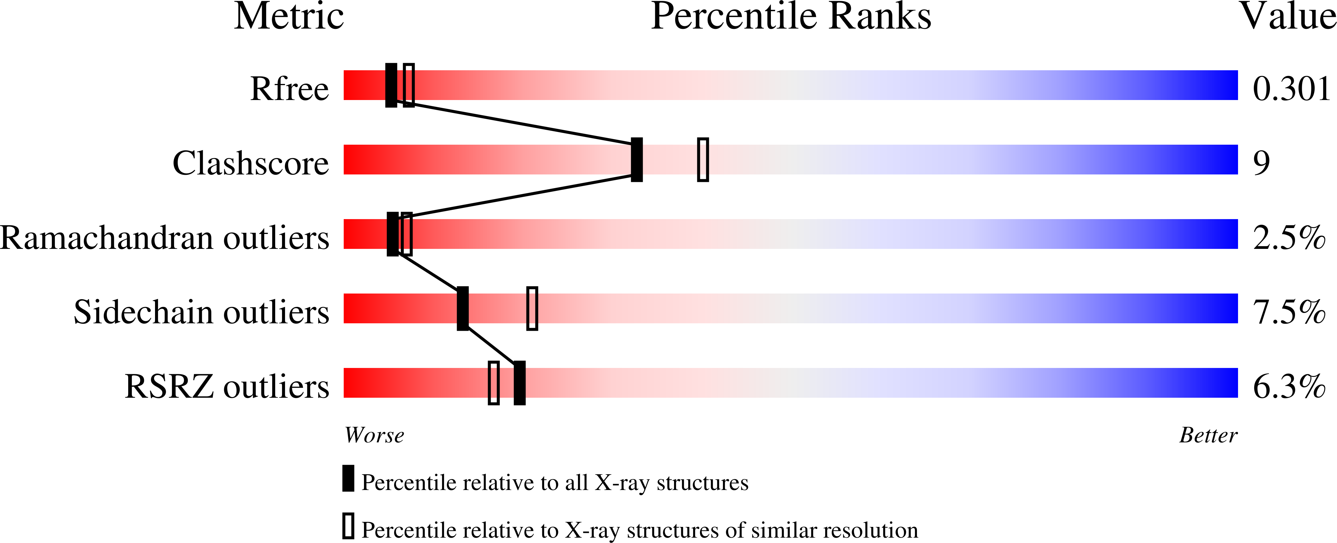
R-Value Free:
0.29
R-Value Work:
0.23
R-Value Observed:
0.24
Space Group:
C 1 2 1