Deposition Date
2006-06-15
Release Date
2007-05-01
Last Version Date
2023-10-25
Entry Detail
PDB ID:
2HC4
Keywords:
Title:
Crystal structure of the LBD of VDR of Danio rerio in complex with calcitriol
Biological Source:
Source Organism(s):
Danio rerio (Taxon ID: 7955)
Homo sapiens (Taxon ID: 9606)
Homo sapiens (Taxon ID: 9606)
Expression System(s):
Method Details:
Experimental Method:
Resolution:
2.20 Å
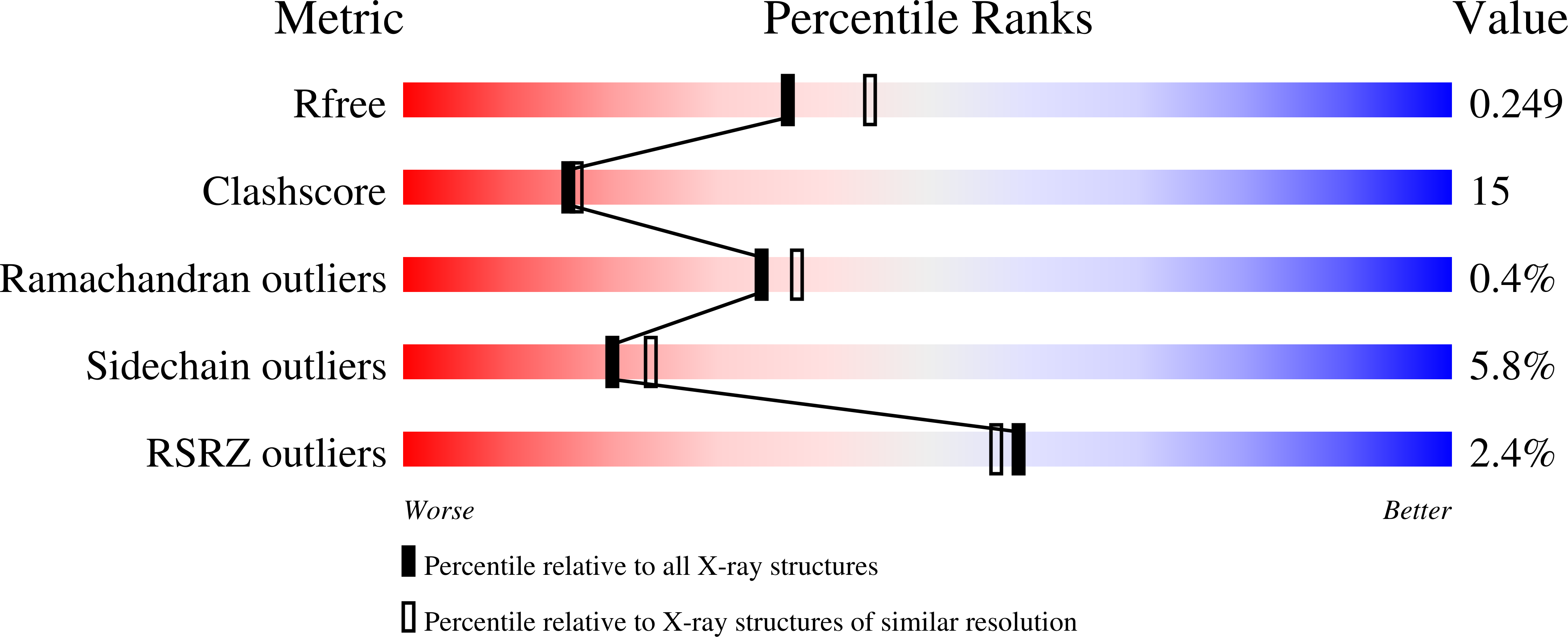
R-Value Free:
0.25
R-Value Work:
0.22
R-Value Observed:
0.22
Space Group:
P 65 2 2