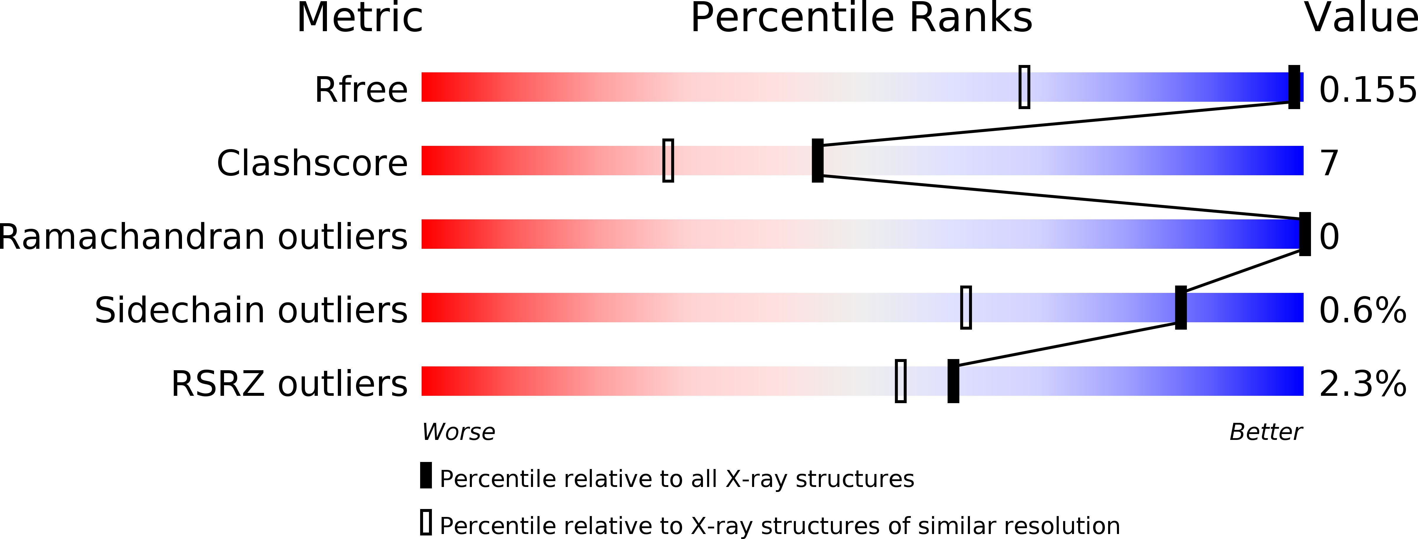
2HBW image
Deposition Date 2006-06-14
Release Date 2006-08-08
Last Version Date 2024-11-20
Entry Detail
PDB ID:
2HBW
Keywords:
Title:
Crystal structure of a putative endopeptidase (ava_3396) from anabaena variabilis atcc 29413 at 1.05 A resolution
Biological Source:
Source Organism:
Host Organism:
Method Details:
Experimental Method:
Resolution:
1.05 Å
R-Value Free:
0.14
R-Value Work:
0.12
R-Value Observed:
0.12
Space Group:
P 21 21 2
Macromolecular Entities
Polymer Type:polypeptide(L)
Molecule:NLP/P60 protein
Gene (Uniprot):Ava_3396
Chain IDs:A
Chain Length:235
Number of Molecules:1
Biological Source:Anabaena variabilis
Modified Residue
Compound ID Chain ID Parent Comp ID Details 2D Image
MSE A MET SELENOMETHIONINE
Primary Citation

Abstact

The crystal structures of two homologous endopeptidases from cyanobacteria Anabaena variabilis and Nostoc punctiforme were determined at 1.05 and 1.60 A resolution, respectively, and contain a bacterial SH3-like domain (SH3b) and a ubiquitous cell-wall-associated NlpC/P60 (or CHAP) cysteine peptidase domain. The NlpC/P60 domain is a primitive, papain-like peptidase in the CA clan of cysteine peptidases with a Cys126/His176/His188 catalytic triad and a conserved catalytic core. We deduced from structure and sequence analysis, and then experimentally, that these two proteins act as gamma-D-glutamyl-L-diamino acid endopeptidases (EC 3.4.22.-). The active site is located near the interface between the SH3b and NlpC/P60 domains, where the SH3b domain may help define substrate specificity, instead of functioning as a targeting domain, so that only muropeptides with an N-terminal L-alanine can bind to the active site.

Legend

Protein

Chemical

Disease

Primary Citation of related structures