Deposition Date
2006-06-14
Release Date
2007-05-01
Last Version Date
2023-10-25
Entry Detail
PDB ID:
2HBH
Keywords:
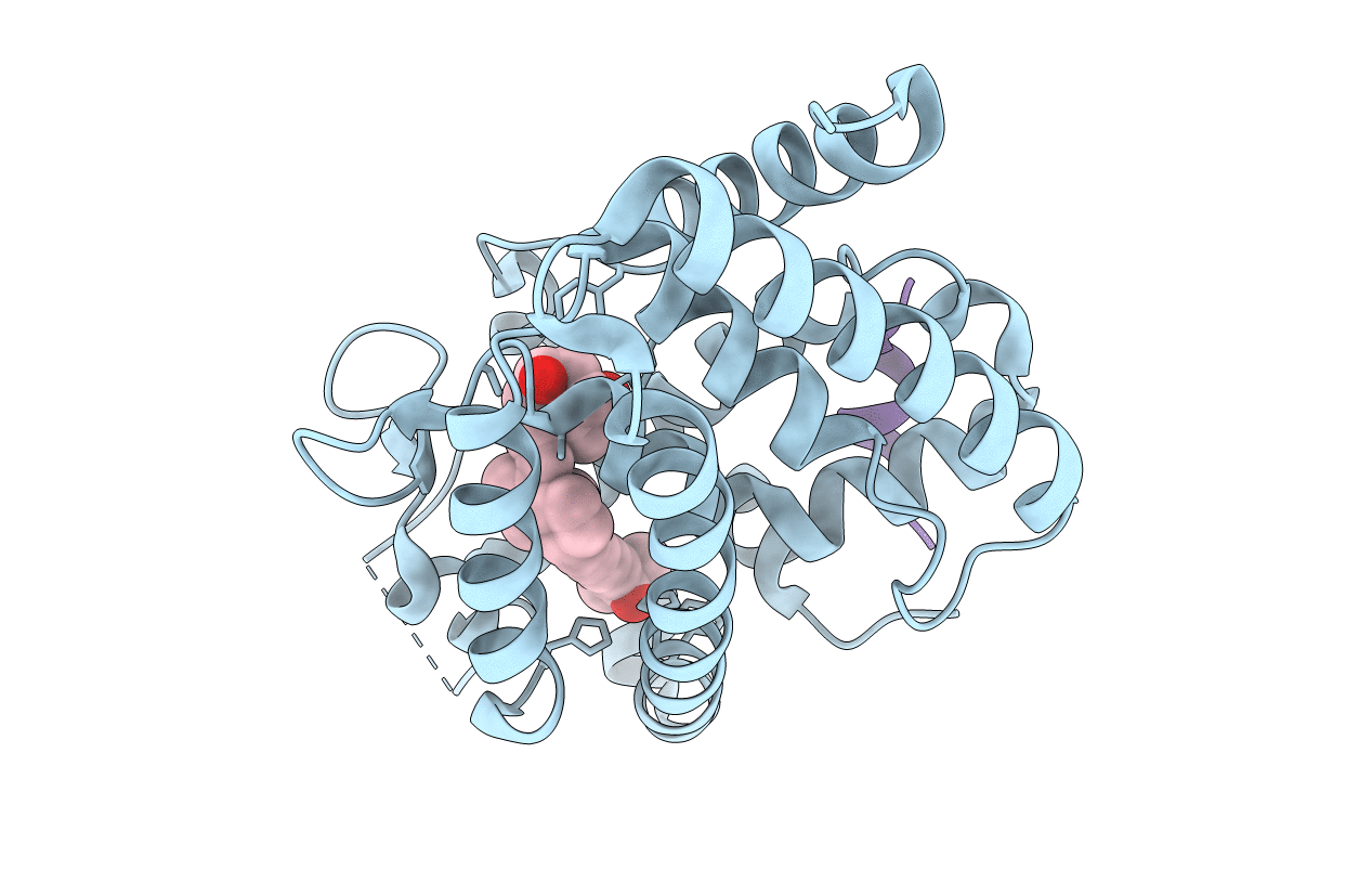
Title:
Crystal structure of Vitamin D nuclear receptor ligand binding domain bound to a locked side-chain analog of calcitriol and SRC-1 peptide
Biological Source:
Source Organism(s):
Danio rerio (Taxon ID: 7955)
Homo sapiens (Taxon ID: 9606)
Homo sapiens (Taxon ID: 9606)
Expression System(s):
Method Details:
Experimental Method:
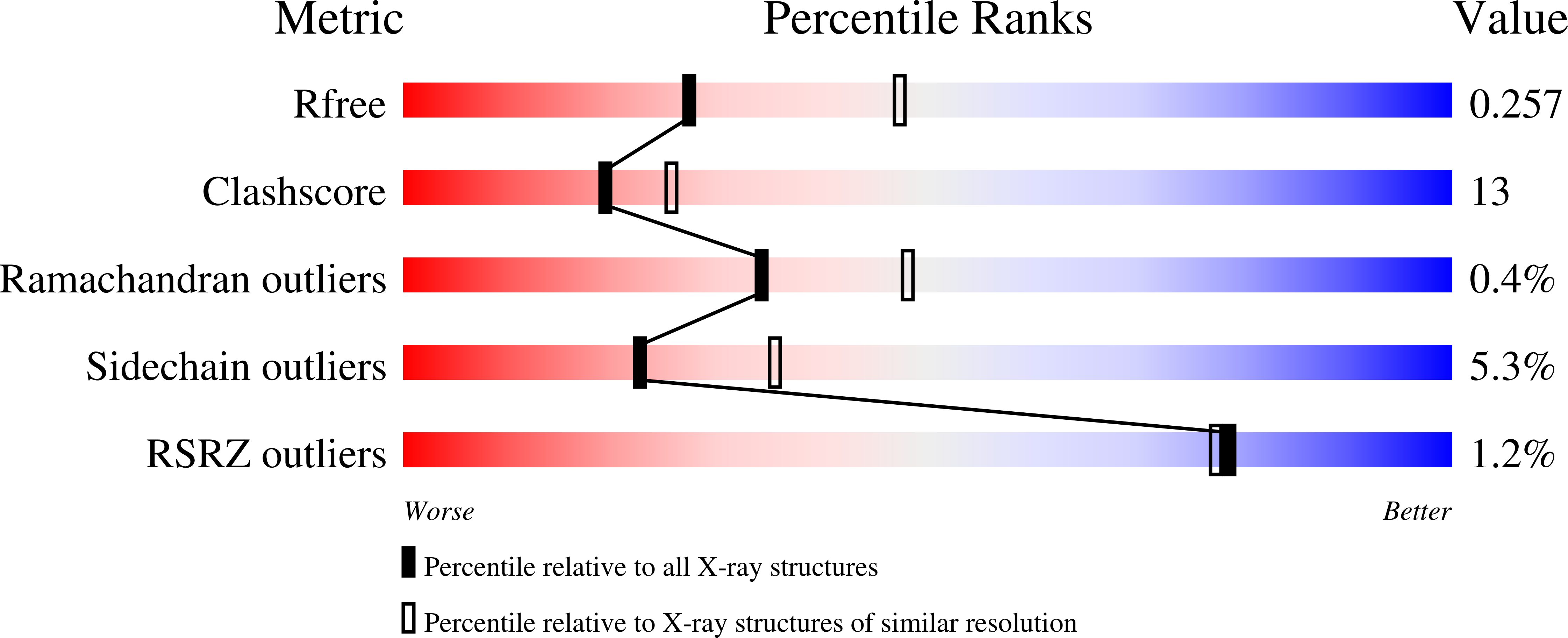
Resolution:
2.65 Å
R-Value Free:
0.26
R-Value Work:
0.20
R-Value Observed:
0.20
Space Group:
P 65 2 2