2HB7 image
Entry Detail
PDB ID:
2HB7
Keywords:
Title:
Crystal structure of VDR LBD in complex with 2alpha(3-hydroxy-1-propyl) calcitriol
Biological Source:
Source Organism:
PDB Version:
Deposition Date:
2006-06-14
Release Date:
2006-08-29
Method Details:
Experimental Method:
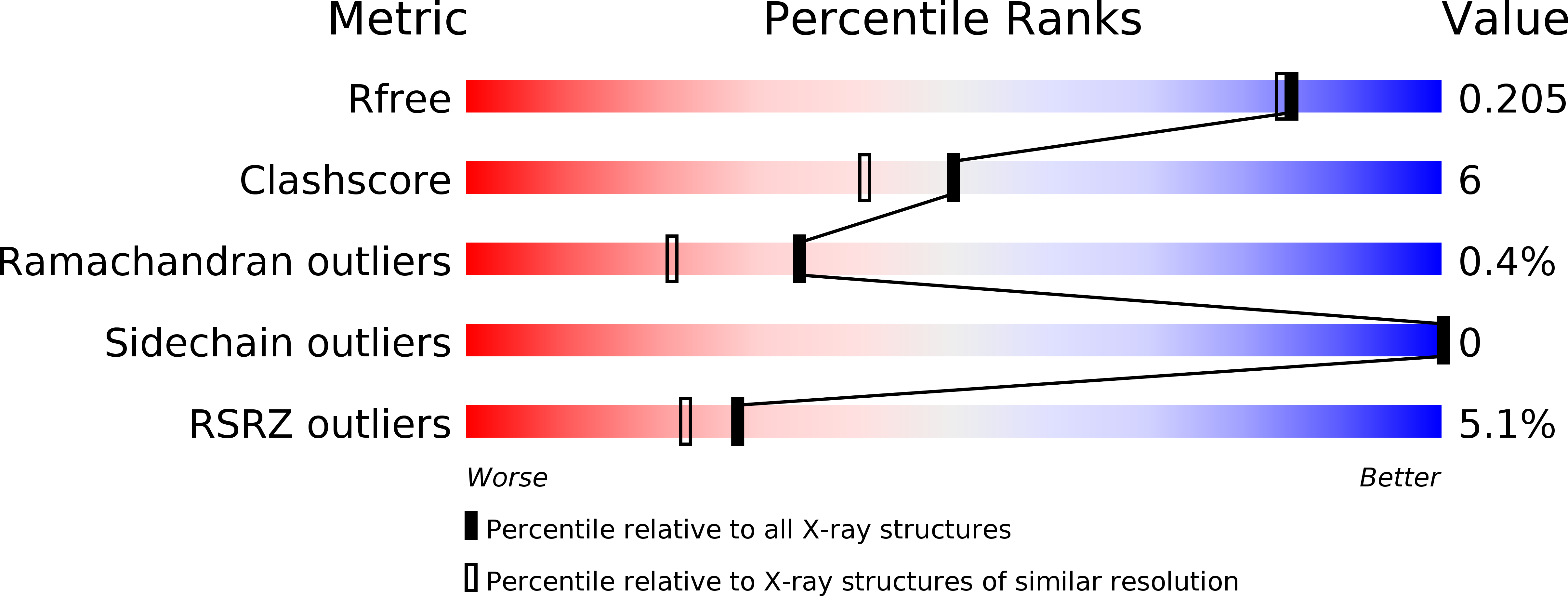
Resolution:
1.80 Å
R-Value Free:
0.20
R-Value Work:
0.18
R-Value Observed:
0.18
Space Group:
P 21 21 21
Macromolecular Entities
Polymer Type:polypeptide(L)
Description:Vitamin D3 receptor
Chain IDs:A
Chain Length:263
Number of Molecules:1
Biological Source:Homo sapiens
Ligand Molecules
Primary Citation
Probing a Water Channel near the A-Ring of Receptor-Bound 1alpha,25-Dihydroxyvitamin D3 with Selected 2alpha-Substituted Analogues
J.Med.Chem. 49 5199 5205 (2006)
PMID: 16913708 DOI: 10.1021/jm0604070

Abstact

The crystal structure of the vitamin D receptor (VDR) in complex with 1 alpha,25(OH)2D3 revealed the presence of several water molecules near the A-ring linking the ligand C-2 position to the protein surface. Here, we report the crystal structures of the human VDR ligand binding domain bound to selected C-2 alpha substituted analogues, namely, methyl, propyl, propoxy, hydroxypropyl, and hydroxypropoxy. These specific replacements do not modify the structure of the protein or the ligand, but with the exception of the methyl substituent, all analogues affect the presence and/or the location of the above water molecules. The integrity of the channel interactions and specific C-2 alpha analogue directed additional interactions correlate with the binding affinity of the ligands. In contrast, the resulting loss or gain of H-bonds does not reflect the magnitude of HL60 cell differentiation. Our overall findings highlight a rational approach to the design of more potent ligands by building in features revealed in the crystal structures.

Legend

Protein

Chemical

Disease

Primary Citation of related structures