2HB5 image
Deposition Date 2006-06-13
Release Date 2006-08-29
Last Version Date 2024-10-16
Entry Detail
PDB ID:
2HB5
Keywords:
Title:
Crystal Structure of the Moloney Murine Leukemia Virus RNase H Domain
Biological Source:
Source Organism:
Host Organism:
Method Details:
Experimental Method:
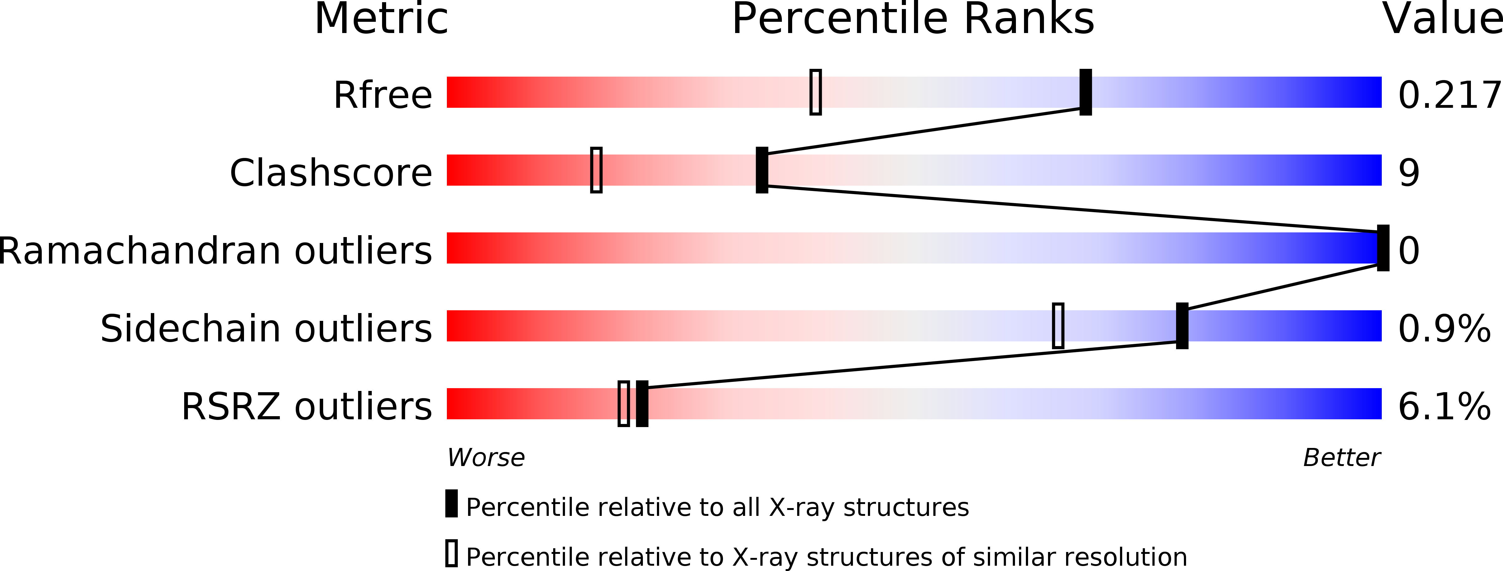
Resolution:
1.59 Å
R-Value Free:
0.22
R-Value Work:
0.19
Space Group:
P 1
Macromolecular Entities
Structures with similar UniProt ID
Protein Blast
Polymer Type:polypeptide(L)
Molecule:Reverse transcriptase/ribonuclease H
Gene (Uniprot):gag-pol
Chain IDs:A
Chain Length:164
Number of Molecules:1
Biological Source:Moloney murine leukemia virus
Modified Residue
Compound ID Chain ID Parent Comp ID Details 2D Image
MSE A MET SELENOMETHIONINE
Primary Citation
Crystal Structure of the Moloney Murine Leukemia Virus RNase H Domain.
J.Virol. 80 8379 8389 (2006)
PMID: 16912289 DOI: 10.1128/JVI.00750-06

Abstact

A crystallographic study of the Moloney murine leukemia virus (Mo-MLV) RNase H domain was performed to provide information about its structure and mechanism of action. These efforts resulted in the crystallization of a mutant Mo-MLV RNase H lacking the putative helix C (DeltaC). The 1.6-Angstroms resolution structure resembles the known structures of the human immunodeficiency virus type 1 (HIV-1) and Escherichia coli RNase H. The structure revealed the coordination of a magnesium ion within the catalytic core comprised of the highly conserved acidic residues D524, E562, and D583. Surface charge mapping of the Mo-MLV structure revealed a high density of basic charges on one side of the enzyme. Using a model of the Mo-MLV structure superimposed upon a structure of HIV-1 reverse transcriptase bound to an RNA/DNA hybrid substrate, Mo-MLV RNase H secondary structures and individual amino acids were examined for their potential roles in binding substrate. Identified regions included Mo-MLV RNase H beta1-beta2, alphaA, and alphaB and residues from alphaB to alphaD and its following loop. Most of the identified substrate-binding residues corresponded with residues directly binding nucleotides in an RNase H from Bacillus halodurans as observed in a cocrystal structure with RNA/DNA. Finally, superimposition of RNases H of Mo-MLV, E. coli, and HIV-1 revealed that a loop of the HIV-1 connection domain resides within the same region of the Mo-MLV and E. coli C-helix. The HIV-1 connection domain may serve to recognize and bind the RNA/DNA substrate major groove.

Legend

Protein

Chemical

Disease

Primary Citation of related structures