2HB3 image
Deposition Date 2006-06-13
Release Date 2006-08-29
Last Version Date 2024-02-14
Entry Detail
PDB ID:
2HB3
Keywords:
Title:
Wild-type HIV-1 Protease in complex with potent inhibitor GRL06579
Biological Source:
Source Organism:
Host Organism:
Method Details:
Experimental Method:
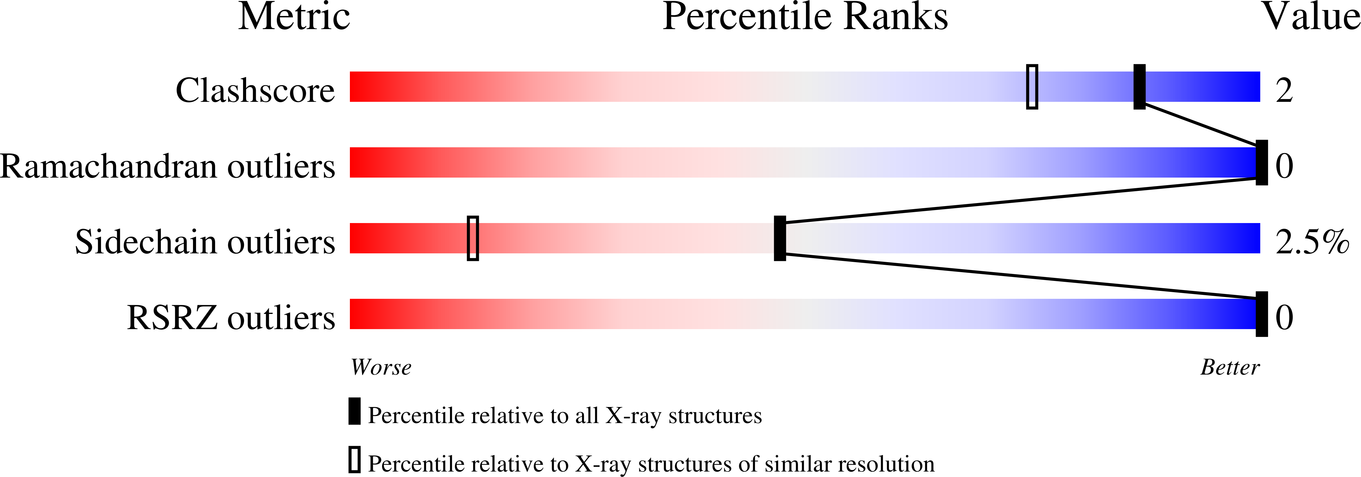
Resolution:
1.35 Å
R-Value Free:
0.19
R-Value Work:
0.14
R-Value Observed:
0.14
Space Group:
P 21 21 2
Macromolecular Entities
Polymer Type:polypeptide(L)
Molecule:Protease
Mutations:Q7K, L33I, L63I, C67A,C95A
Chain IDs:A, B
Chain Length:99
Number of Molecules:2
Biological Source:Human immunodeficiency virus 1
Primary Citation
Structure-Based Design of Novel HIV-1 Protease Inhibitors To Combat Drug Resistance.
J.Med.Chem. 49 5252 5261 (2006)
PMID: 16913714 DOI: 10.1021/jm060561m

Abstact

Structure-based design and synthesis of novel HIV protease inhibitors are described. The inhibitors are designed specifically to interact with the backbone of HIV protease active site to combat drug resistance. Inhibitor 3 has exhibited exceedingly potent enzyme inhibitory and antiviral potency. Furthermore, this inhibitor maintains impressive potency against a wide spectrum of HIV including a variety of multi-PI-resistant clinical strains. The inhibitors incorporated a stereochemically defined 5-hexahydrocyclopenta[b]furanyl urethane as the P2-ligand into the (R)-(hydroxyethylamino)sulfonamide isostere. Optically active (3aS,5R,6aR)-5-hydroxy-hexahydrocyclopenta[b]furan was prepared by an enzymatic asymmetrization of meso-diacetate with acetyl cholinesterase, radical cyclization, and Lewis acid-catalyzed anomeric reduction as the key steps. A protein-ligand X-ray crystal structure of inhibitor 3-bound HIV-1 protease (1.35 A resolution) revealed extensive interactions in the HIV protease active site including strong hydrogen bonding interactions with the backbone. This design strategy may lead to novel inhibitors that can combat drug resistance.

Legend

Protein

Chemical

Disease

Primary Citation of related structures