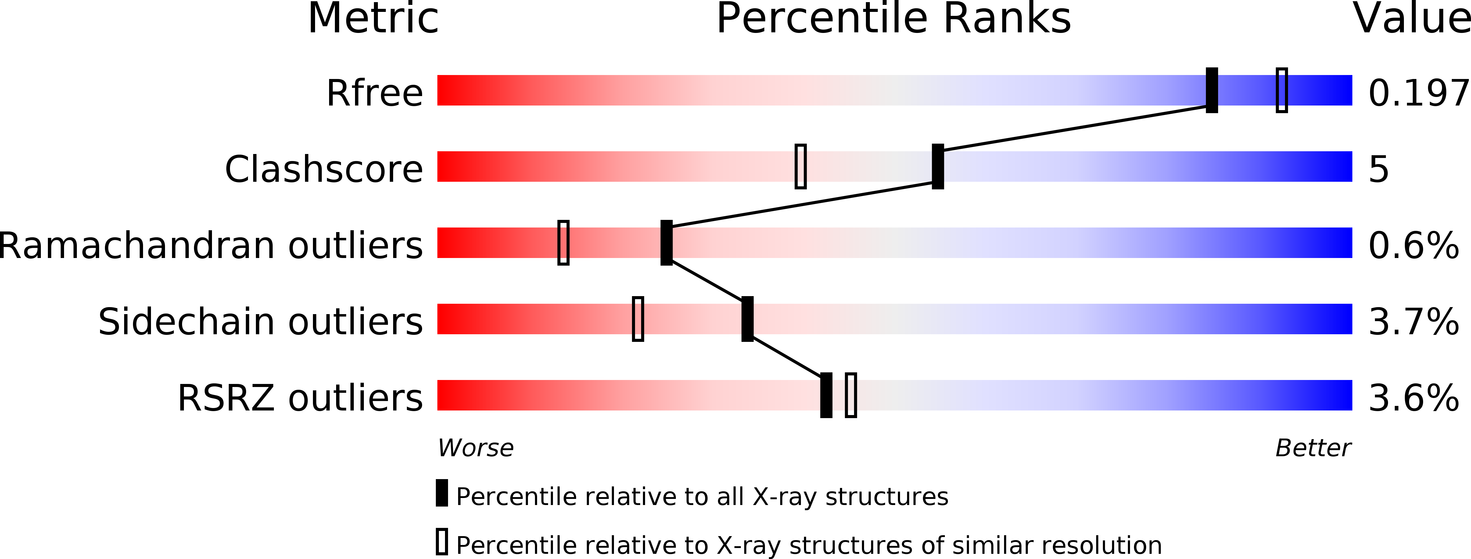
2HAQ image
Deposition Date 2006-06-13
Release Date 2006-06-20
Last Version Date 2023-08-30
Entry Detail
PDB ID:
2HAQ
Keywords:
Title:
Crystal Structure of Cyclophilin A from Leishmania Donovani
Biological Source:
Source Organism:
Host Organism:
Method Details:
Experimental Method:
Resolution:
1.97 Å
R-Value Free:
0.19
R-Value Work:
0.17
R-Value Observed:
0.17
Space Group:
P 43 21 2
Macromolecular Entities
Structures with similar UniProt ID
Protein Blast
Polymer Type:polypeptide(L)
Molecule:Cyclophilin
Gene (Uniprot):CYP
Chain IDs:A
Chain Length:172
Number of Molecules:1
Biological Source:Leishmania donovani
Primary Citation
Structure of cyclophilin from Leishmania donovani at 1.97 A resolution.
Acta Crystallogr.,Sect.F 63 60 64 (2007)
PMID: 17277440 DOI: 10.1107/S1744309106056351

Abstact

The crystal structure of cyclophilin from Leishmania donovani (LdCyp) has been determined and refined at 1.97 A resolution to a crystallographic R factor of 0.178 (R(free) = 0.197). The structure was solved by molecular replacement using cyclophilin from Trypanosoma cruzi as the search model. LdCyp exhibits complete structural conservation of the cyclosporin-binding site with respect to the homologous human protein, as anticipated from LdCyp-cyclosporin binding studies. Comparisons with other cyclophilins show deviations primarily in the loop regions. The solvent structure encompassing the molecule has also been analyzed in some detail.

Legend

Protein

Chemical

Disease

Primary Citation of related structures