Deposition Date
2006-06-12
Release Date
2006-08-08
Last Version Date
2024-11-06
Entry Detail
PDB ID:
2HAG
Keywords:
Title:
Crystal structure of a putative dyp-type peroxidase protein (so_0740) from shewanella oneidensis at 2.75 A resolution
Biological Source:
Source Organism(s):
Shewanella oneidensis (Taxon ID: 70863)
Expression System(s):
Method Details:
Experimental Method:
Resolution:
2.75 Å
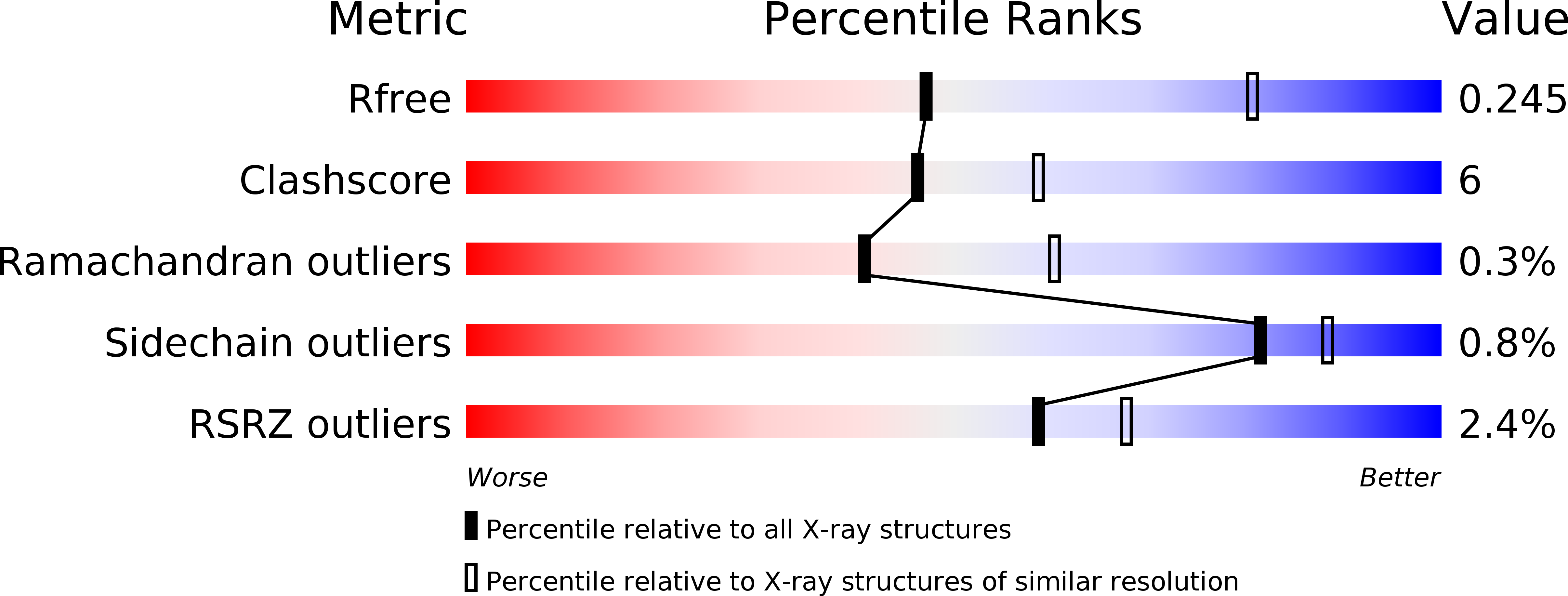
R-Value Free:
0.22
R-Value Work:
0.19
R-Value Observed:
0.19
Space Group:
P 43 21 2