2HA6 image
Entry Detail
PDB ID:
2HA6
Keywords:
Title:
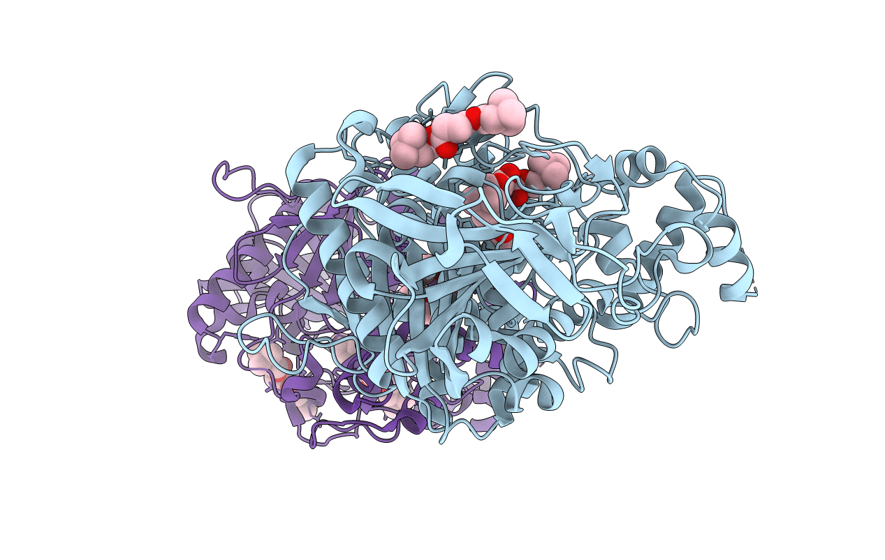
Crystal structure of mutant S203A of mouse acetylcholinesterase complexed with succinylcholine
Biological Source:
Source Organism:
Host Organism:
PDB Version:
Deposition Date:
2006-06-12
Release Date:
2006-07-18
Method Details:
Experimental Method:
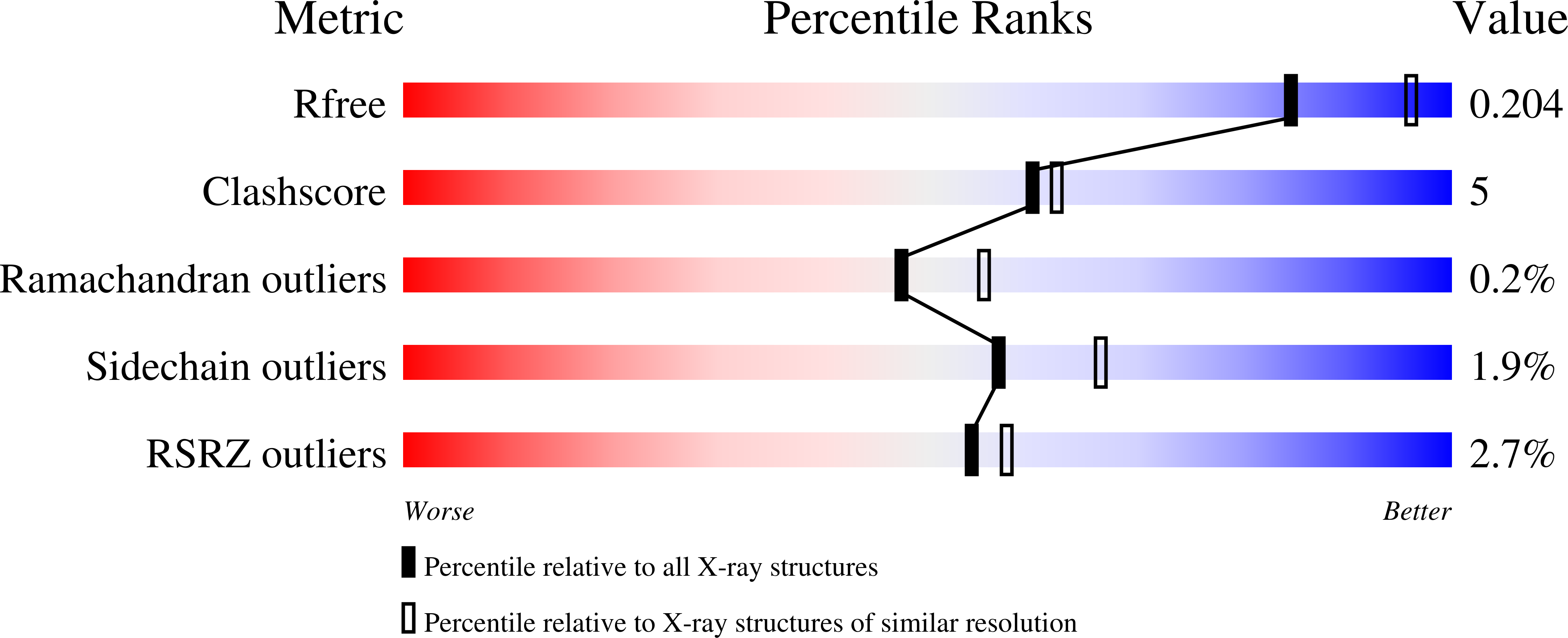
Resolution:
2.25 Å
R-Value Free:
0.19
R-Value Work:
0.17
R-Value Observed:
0.17
Space Group:
P 21 21 21
Macromolecular Entities
Polymer Type:polypeptide(L)
Description:Acetylcholinesterase
Mutations:S203A
Chain IDs:A, B
Chain Length:543
Number of Molecules:2
Biological Source:Mus musculus
Primary Citation
Substrate and product trafficking through the active center gorge of acetylcholinesterase analyzed by crystallography and equilibrium binding
J.Biol.Chem. 281 29256 29267 (2006)
PMID: 16837465 DOI: 10.1074/jbc.M603018200

Abstact

Hydrolysis of acetylcholine catalyzed by acetylcholinesterase (AChE), one of the most efficient enzymes in nature, occurs at the base of a deep and narrow active center gorge. At the entrance of the gorge, the peripheral anionic site provides a binding locus for allosteric ligands, including substrates. To date, no structural information on substrate entry to the active center from the peripheral site of AChE or its subsequent egress has been reported. Complementary crystal structures of mouse AChE and an inactive mouse AChE mutant with a substituted catalytic serine (S203A), in various complexes with four substrates (acetylcholine, acetylthiocholine, succinyldicholine, and butyrylthiocholine), two non-hydrolyzable substrate analogues (m-(N,N,N-trimethylammonio)-trifluoroacetophenone and 4-ketoamyltrimethylammonium), and one reaction product (choline) were solved in the 2.05-2.65-A resolution range. These structures, supported by binding and inhibition data obtained on the same complexes, reveal the successive positions and orientations of the substrates bound to the peripheral site and proceeding within the gorge toward the active site, the conformations of the presumed transition state for acylation and the acyl-enzyme intermediate, and the positions and orientations of the dissociating and egressing products. Moreover, the structures of the AChE mutant in complexes with acetylthiocholine and succinyldicholine reveal additional substrate binding sites on the enzyme surface, distal to the gorge entry. Hence, we provide a comprehensive set of structural snapshots of the steps leading to the intermediates of catalysis and the potential regulation by substrate binding to various allosteric sites at the enzyme surface.

Legend

Protein

Chemical

Disease

Primary Citation of related structures