2H8A image
Entry Detail
PDB ID:
2H8A
Keywords:
Title:
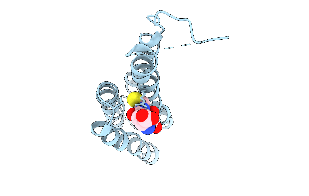
Structure of Microsomal Glutathione Transferase 1 in Complex with Glutathione
Biological Source:
Source Organism:
PDB Version:
Deposition Date:
2006-06-07
Release Date:
2007-05-22
Method Details:
Experimental Method:
Resolution:
3.20 Å
R-Value Free:
0.37
R-Value Work:
0.33
R-Value Observed:
0.34
Space Group:
P 6
Macromolecular Entities
Polymer Type:polypeptide(L)
Description:Microsomal glutathione S-transferase 1
Chain IDs:A
Chain Length:154
Number of Molecules:1
Biological Source:Rattus norvegicus
Ligand Molecules
Primary Citation
Structural basis for detoxification and oxidative stress protection in membranes.
J.Mol.Biol. 360 934 945 (2006)
PMID: 16806268 DOI: 10.1016/j.jmb.2006.05.056

Abstact

Synthesis of mediators of fever, pain and inflammation as well as protection against reactive molecules and oxidative stress is a hallmark of the MAPEG superfamily (membrane associated proteins in eicosanoid and glutathione metabolism). The structure of a MAPEG member, rat microsomal glutathione transferase 1, at 3.2 A resolution, solved here in complex with glutathione by electron crystallography, defines the active site location and a cytosolic domain involved in enzyme activation. The glutathione binding site is found to be different from that of the canonical soluble glutathione transferases. The architecture of the homotrimer supports a catalytic mechanism involving subunit interactions and reveals both cytosolic and membraneous substrate entry sites, providing a rationale for the membrane location of the enzyme.

Legend

Protein

Chemical

Disease

Primary Citation of related structures