2H7G image
Deposition Date 2006-06-02
Release Date 2006-08-15
Last Version Date 2024-02-14
Entry Detail
PDB ID:
2H7G
Keywords:
Title:
Structure of variola topoisomerase non-covalently bound to DNA
Biological Source:
Source Organism:
Variola virus (Taxon ID: 10255)
Method Details:
Experimental Method:
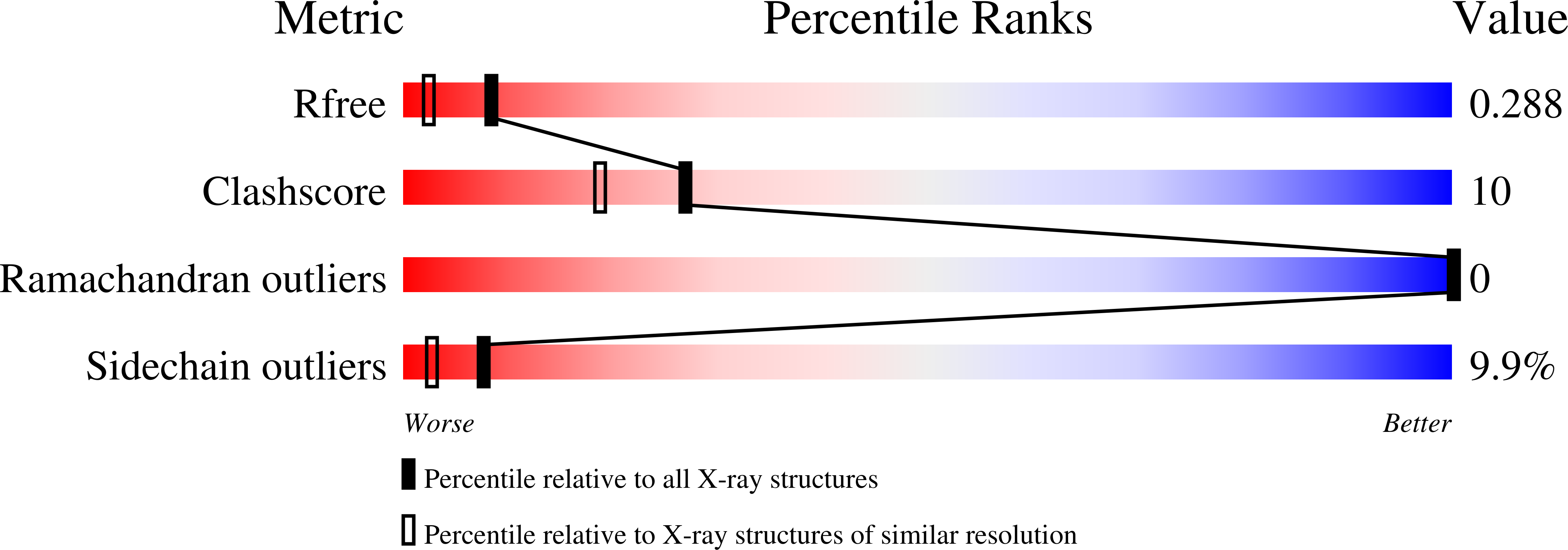
Resolution:
1.90 Å
R-Value Free:
0.24
R-Value Work:
0.19
R-Value Observed:
0.19
Space Group:
C 2 2 21
Macromolecular Entities
Structures with similar UniProt ID
Protein Blast
Polymer Type:polypeptide(L)
Molecule:DNA topoisomerase 1
Gene (Uniprot):OPG111
Mutagens:C100S, C211S
Chain IDs:C (auth: X)
Chain Length:314
Number of Molecules:1
Biological Source:Variola virus
Primary Citation
Structural basis for specificity in the poxvirus topoisomerase.
Mol.Cell 23 343 354 (2006)
PMID: 16885024 DOI: 10.1016/j.molcel.2006.06.015

Abstact

Although smallpox has been eradicated from the human population, it is presently feared as a possible agent of bioterrorism. The smallpox virus codes for its own topoisomerase enzyme that differs from its cellular counterpart by requiring a specific DNA sequence for activation of catalysis. Here we present crystal structures of the smallpox virus topoisomerase enzyme bound both covalently and noncovalently to a specific DNA sequence. These structures reveal the basis for site-specific DNA recognition, and they explain how catalysis is likely activated by formation of a specific enzyme-DNA interface. Unexpectedly, the poxvirus enzyme uses a major groove binding alpha helix that is not present in the human enzyme to recognize part of the core recognition sequence and activate the enzyme for catalysis. The topoisomerase-DNA complex structures also provide a three-dimensional framework that may facilitate the rational design of therapeutic agents to treat poxvirus infections.

Legend

Protein

Chemical

Disease

Primary Citation of related structures