Deposition Date
2006-05-30
Release Date
2006-09-19
Last Version Date
2023-11-15
Entry Detail
PDB ID:
2H65
Keywords:
Title:
Crystal strusture of caspase-3 with inhibitor Ac-VDVAD-Cho
Biological Source:
Source Organism(s):
Homo sapiens (Taxon ID: 9606)
Expression System(s):
Method Details:
Experimental Method:
Resolution:
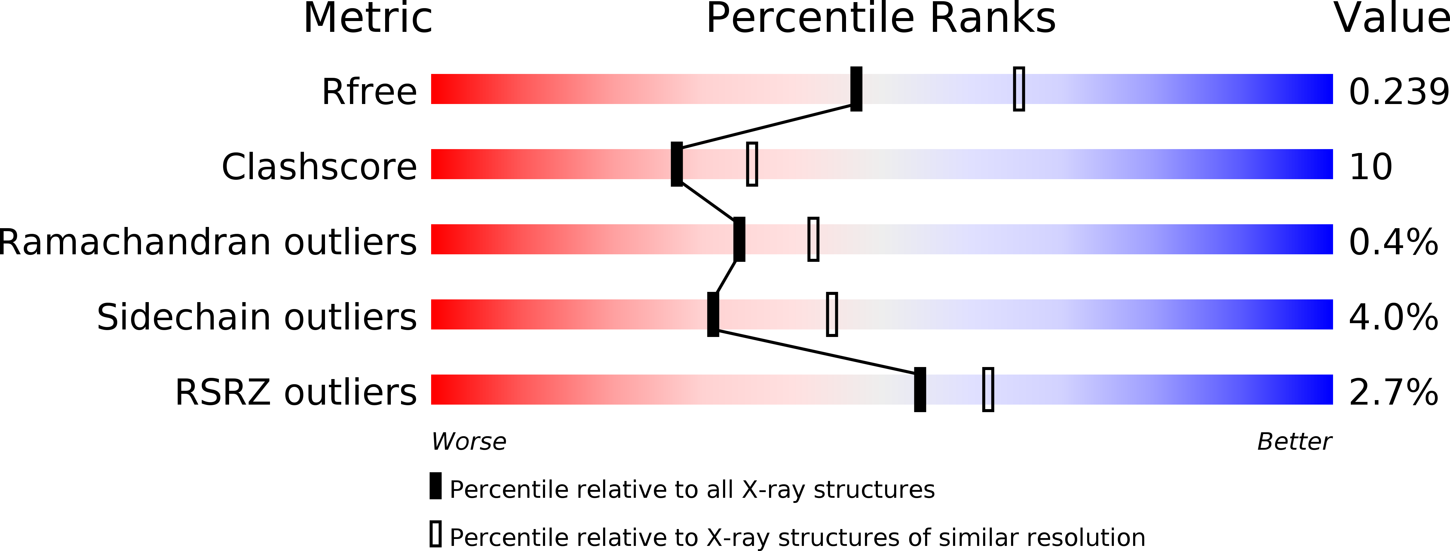
2.30 Å
R-Value Free:
0.24
R-Value Work:
0.20
R-Value Observed:
0.19
Space Group:
P 1 21 1