2H4T image
Deposition Date 2006-05-25
Release Date 2006-07-25
Last Version Date 2024-02-14
Entry Detail
PDB ID:
2H4T
Keywords:
Title:
Crystal structure of rat carnitine palmitoyltransferase II
Biological Source:
Source Organism:
Host Organism:
Method Details:
Experimental Method:
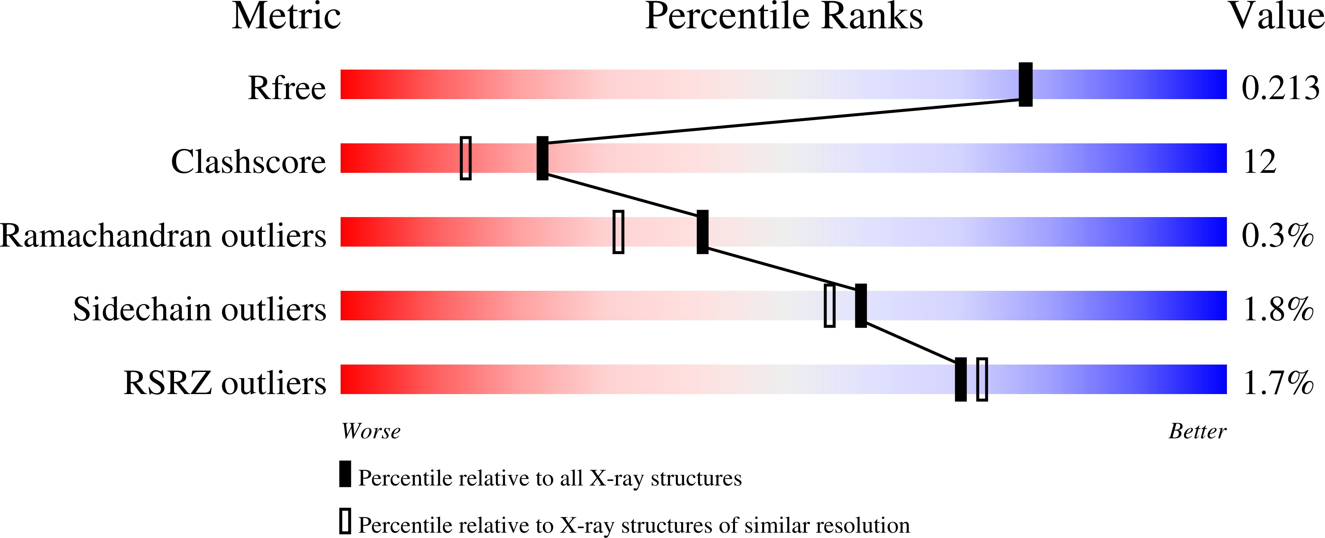
Resolution:
1.90 Å
R-Value Free:
0.22
R-Value Work:
0.18
R-Value Observed:
0.18
Space Group:
P 1
Macromolecular Entities
Polymer Type:polypeptide(L)
Molecule:Carnitine O-palmitoyltransferase II, mitochondrial
Gene (Uniprot):Cpt2
Mutations:A547T
Chain IDs:A, B
Chain Length:626
Number of Molecules:2
Biological Source:Rattus norvegicus
Ligand Molecules
Primary Citation
Crystal structure of rat carnitine palmitoyltransferase II (CPT-II).
Biochem.Biophys.Res.Commun. 346 974 980 (2006)
PMID: 16781677 DOI: 10.1016/j.bbrc.2006.06.006

Abstact

Carnitine palmitoyltransferase II (CPT-II) has a crucial role in the beta-oxidation of long-chain fatty acids in mitochondria. We report here the crystal structure of rat CPT-II at 1.9A resolution. The overall structure shares strong similarity to those of short- and medium-chain carnitine acyltransferases, although detailed structural differences in the active site region have a significant impact on the substrate selectivity of CPT-II. Three aliphatic chains, possibly from a detergent that is used for the crystallization, were found in the structure. Two of them are located in the carnitine and CoA binding sites, respectively. The third aliphatic chain may mimic the long-chain acyl group in the substrate of CPT-II. The binding site for this aliphatic chain does not exist in the short- and medium-chain carnitine acyltransferases, due to conformational differences among the enzymes. A unique insert in CPT-II is positioned on the surface of the enzyme, with a highly hydrophobic surface. It is likely that this surface patch mediates the association of CPT-II with the inner membrane of the mitochondria.

Legend

Protein

Chemical

Disease

Primary Citation of related structures