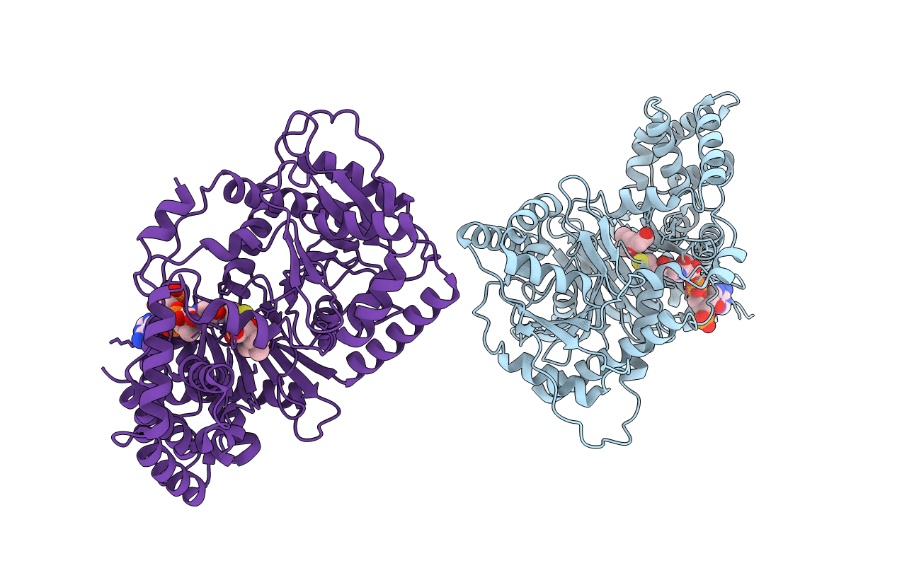
2H3U image
Entry Detail
PDB ID:
2H3U
Keywords:
Title:
Crystal structure of murine carnitine acetyltransferase in complex with carnitine and CoA
Biological Source:
Source Organism:
Host Organism:
PDB Version:
Deposition Date:
2006-05-23
Release Date:
2006-08-01
Method Details:
Experimental Method:
Resolution:
1.90 Å
R-Value Free:
0.23
R-Value Work:
0.19
R-Value Observed:
0.19
Space Group:
C 1 2 1
Macromolecular Entities
Polymer Type:polypeptide(L)
Description:carnitine acetyltransferase
Chain IDs:A, B
Chain Length:599
Number of Molecules:2
Biological Source:Mus musculus
Primary Citation
Crystal structures of murine carnitine acetyltransferase in ternary complexes with its substrates
J.Biol.Chem. 281 28480 28487 (2006)
PMID: 16870616 DOI: 10.1074/jbc.M602622200

Abstact

Carnitine acyltransferases catalyze the reversible exchange of acyl groups between coenzyme A (CoA) and carnitine. They have important roles in many cellular processes, especially the oxidation of long-chain fatty acids in the mitochondria for energy production, and are attractive targets for drug discovery against diabetes and obesity. To help define in molecular detail the catalytic mechanism of these enzymes, we report here the high resolution crystal structure of wild-type murine carnitine acetyltransferase (CrAT) in a ternary complex with its substrates acetyl-CoA and carnitine, and the structure of the S554A/M564G double mutant in a ternary complex with the substrates CoA and hexanoylcarnitine. Detailed analyses suggest that these structures may be good mimics for the Michaelis complexes for the forward and reverse reactions of the enzyme, representing the first time that such complexes of CrAT have been studied in molecular detail. The structural information provides significant new insights into the catalytic mechanism of CrAT and possibly carnitine acyltransferases in general.

Legend

Protein

Chemical

Disease

Primary Citation of related structures