2H3G image
Deposition Date 2006-05-22
Release Date 2007-03-20
Last Version Date 2024-02-14
Entry Detail
PDB ID:
2H3G
Title:
Structure of the Type III Pantothenate Kinase (CoaX) from Bacillus Anthracis
Biological Source:
Source Organism:
Host Organism:
Method Details:
Experimental Method:
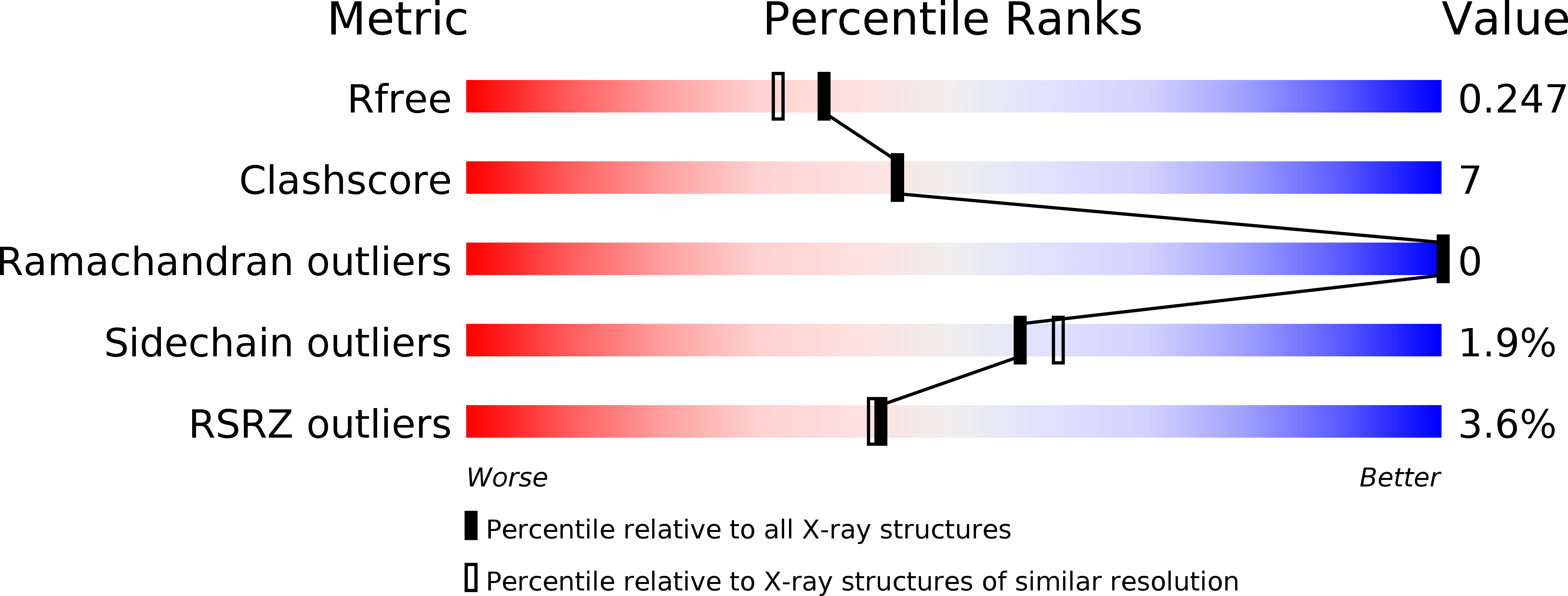
Resolution:
2.00 Å
R-Value Free:
0.25
R-Value Work:
0.21
R-Value Observed:
0.21
Space Group:
P 21 21 2
Macromolecular Entities
Structures with similar UniProt ID
Protein Blast
Polymer Type:polypeptide(L)
Molecule:BIOSYNTHETIC PROTEIN
Chain IDs:A (auth: X)
Chain Length:268
Number of Molecules:1
Biological Source:Bacillus anthracis str.
Ligand Molecules
Primary Citation
Structure of the Type III Pantothenate Kinase from Bacillus anthracis at 2.0 A Resolution: Implications for Coenzyme A-Dependent Redox Biology.
Biochemistry 46 3234 3245 (2007)
PMID: 17323930 DOI: 10.1021/bi062299p

Abstact

Coenzyme A (CoASH) is the major low-molecular weight thiol in Staphylococcus aureus and a number of other bacteria; the crystal structure of the S. aureus coenzyme A-disulfide reductase (CoADR), which maintains the reduced intracellular state of CoASH, has recently been reported [Mallett, T.C., Wallen, J.R., Karplus, P.A., Sakai, H., Tsukihara, T., and Claiborne, A. (2006) Biochemistry 45, 11278-89]. In this report we demonstrate that CoASH is the major thiol in Bacillus anthracis; a bioinformatics analysis indicates that three of the four proteins responsible for the conversion of pantothenate (Pan) to CoASH in Escherichia coli are conserved in B. anthracis. In contrast, a novel type III pantothenate kinase (PanK) catalyzes the first committed step in the biosynthetic pathway in B. anthracis; unlike the E. coli type I PanK, this enzyme is not subject to feedback inhibition by CoASH. The crystal structure of B. anthracis PanK (BaPanK), solved using multiwavelength anomalous dispersion data and refined at a resolution of 2.0 A, demonstrates that BaPanK is a new member of the Acetate and Sugar Kinase/Hsc70/Actin (ASKHA) superfamily. The Pan and ATP substrates have been modeled into the active-site cleft; in addition to providing a clear rationale for the absence of CoASH inhibition, analysis of the Pan-binding pocket has led to the development of two new structure-based motifs (the PAN and INTERFACE motifs). Our analyses also suggest that the type III PanK in the spore-forming B. anthracis plays an essential role in the novel thiol/disulfide redox biology of this category A biodefense pathogen.

Legend

Protein

Chemical

Disease

Primary Citation of related structures