2H30 image
Deposition Date 2006-05-20
Release Date 2006-08-22
Last Version Date 2024-11-13
Entry Detail
PDB ID:
2H30
Keywords:
Title:
Crystal structure of the N-terminal domain of PilB from Neisseria gonorrhoeae
Biological Source:
Source Organism:
Host Organism:
Method Details:
Experimental Method:
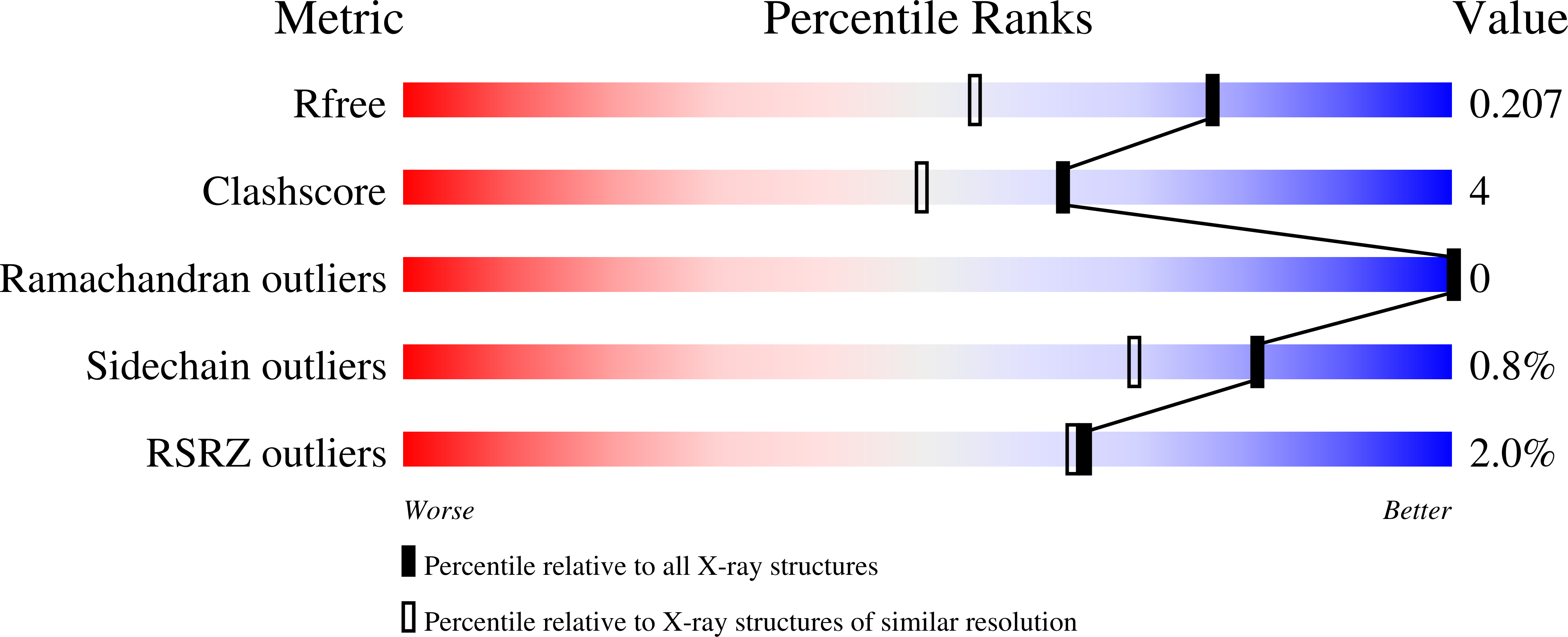
Resolution:
1.60 Å
R-Value Free:
0.20
R-Value Work:
0.18
Space Group:
P 21 21 21
Macromolecular Entities
Structures with similar UniProt ID
Protein Blast
Polymer Type:polypeptide(L)
Molecule:Peptide methionine sulfoxide reductase msrA/msrB
Gene (Uniprot):msrAB
Mutagens:L38M, L41M
Chain IDs:A
Chain Length:164
Number of Molecules:1
Biological Source:Neisseria gonorrhoeae
Modified Residue
Compound ID Chain ID Parent Comp ID Details 2D Image
MSE A MET SELENOMETHIONINE
Primary Citation
The Thioredoxin Domain of Neisseria gonorrhoeae PilB Can Use Electrons from DsbD to Reduce Downstream Methionine Sulfoxide Reductases.
J.Biol.Chem. 281 32668 32675 (2006)
PMID: 16926157 DOI: 10.1074/jbc.M604971200

Abstact

The PilB protein from Neisseria gonorrhoeae is located in the periplasm and made up of three domains. The N-terminal, thioredoxin-like domain (NT domain) is fused to tandem methionine sulfoxide reductase A and B domains (MsrA/B). We show that the alpha domain of Escherichia coli DsbD is able to reduce the oxidized NT domain, which suggests that DsbD in Neisseria can transfer electrons from the cytoplasmic thioredoxin to the periplasm for the reduction of the MsrA/B domains. An analysis of the available complete genomes provides further evidence for this proposition in other bacteria where DsbD/CcdA, Trx, MsrA, and MsrB gene homologs are all located in a gene cluster with a common transcriptional direction. An examination of wild-type PilB and a panel of Cys to Ser mutants of the full-length protein and the individually expressed domains have also shown that the NT domain more efficiently reduces the MsrA/B domains when in the polyprotein context. Within this frame-work there does not appear to be a preference for the NT domain to reduce the proximal MsrA domain over MsrB domain. Finally, we report the 1.6A crystal structure of the NT domain. This structure confirms the presence of a surface loop that makes it different from other membrane-tethered, Trx-like molecules, including TlpA, CcmG, and ResA. Subtle differences are observed in this loop when compared with the Neisseria meningitidis NT domain structure. The data taken together supports the formation of specific NT domain interactions with the MsrA/B domains and its in vivo recycling partner, DsbD.

Legend

Protein

Chemical

Disease

Primary Citation of related structures