
Deposition Date
2006-05-19
Release Date
2006-07-18
Last Version Date
2023-08-30
Entry Detail
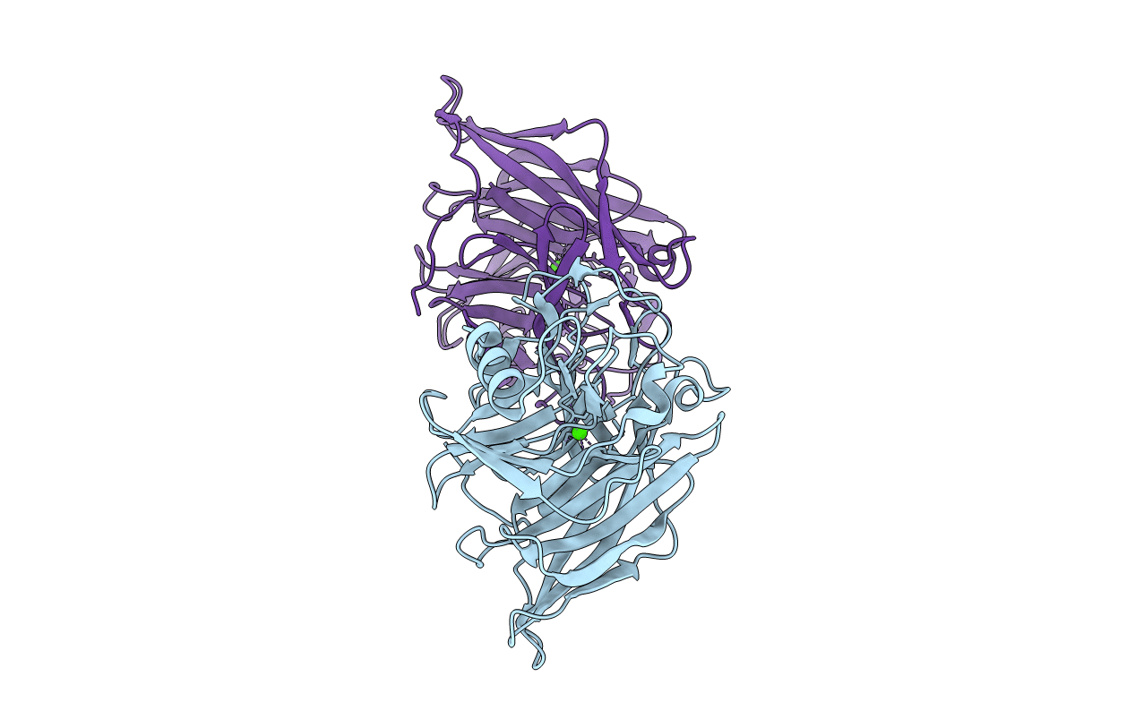
PDB ID:
2H2U
Keywords:
Title:
Crystal structure of the E130Y mutant of human soluble calcium-activated nucleotidase (SCAN) with calcium ion
Biological Source:
Source Organism(s):
Homo sapiens (Taxon ID: 9606)
Expression System(s):
Method Details:
Experimental Method:
Resolution:
2.40 Å
R-Value Free:
0.28
R-Value Work:
0.21
R-Value Observed:
0.22
Space Group:
P 1
