2H1O image
Deposition Date 2006-05-16
Release Date 2006-09-26
Last Version Date 2023-08-30
Entry Detail
PDB ID:
2H1O
Title:
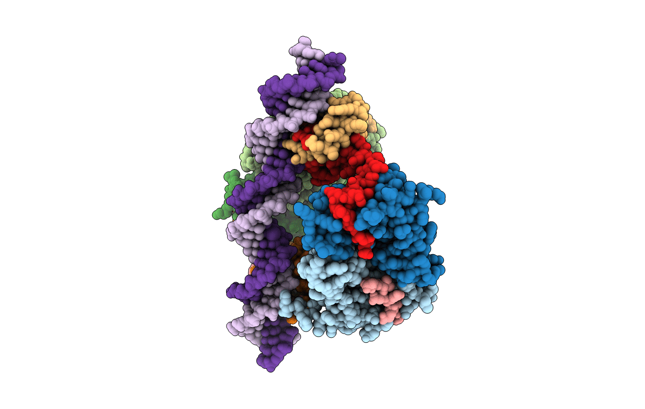
Structure of FitAB bound to IR36 DNA fragment
Biological Source:
Source Organism:
Host Organism:
Method Details:
Experimental Method:
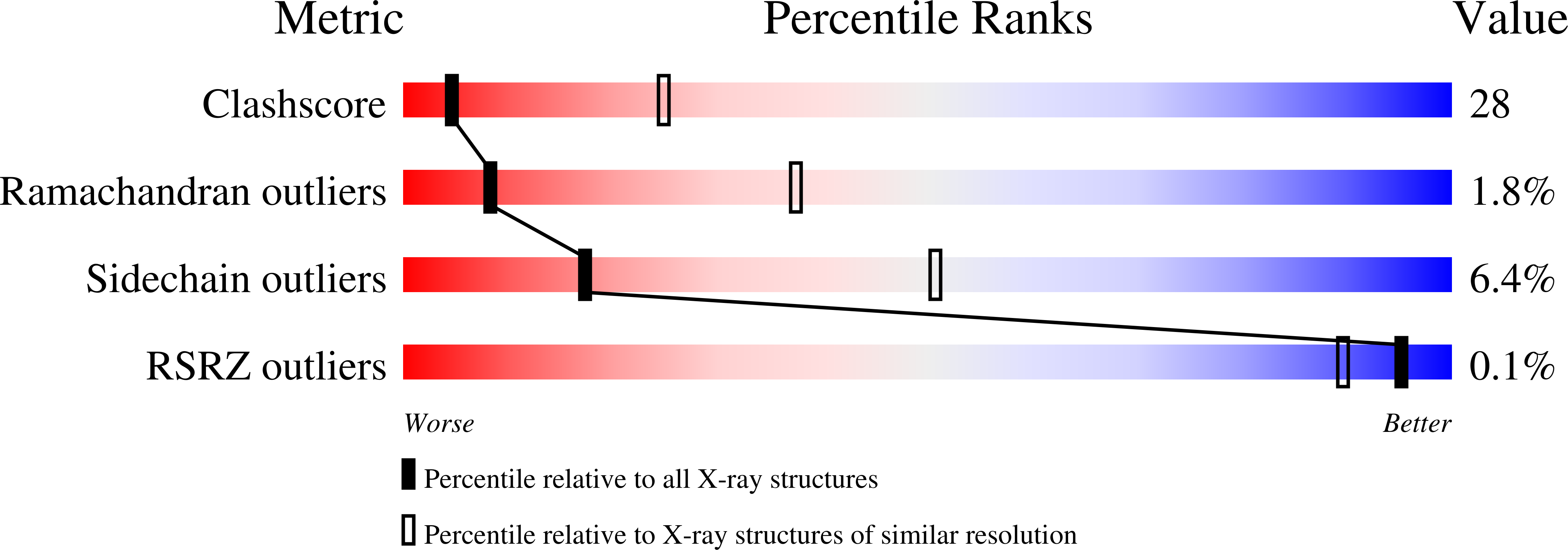
Resolution:
3.00 Å
R-Value Free:
0.26
R-Value Work:
0.21
R-Value Observed:
0.26
Space Group:
P 1 21 1
Macromolecular Entities
Structures with similar UniProt ID
Protein Blast
Polymer Type:polypeptide(L)
Molecule:Trafficking protein B
Chain IDs:C (auth: A), D (auth: B), E (auth: C), F (auth: D)
Chain Length:143
Number of Molecules:4
Biological Source:Neisseria gonorrhoeae
Structures with similar UniProt ID
Protein Blast
Polymer Type:polypeptide(L)
Molecule:Trafficking protein A
Chain IDs:G (auth: E), H (auth: F), I (auth: G), J (auth: H)
Chain Length:68
Number of Molecules:4
Biological Source:Neisseria gonorrhoeae
Polymer Type:polydeoxyribonucleotide
Molecule:IR36-strand 1
Mutagens:iodo
Chain IDs:A (auth: U)
Chain Length:36
Number of Molecules:1
Biological Source:
Polymer Type:polydeoxyribonucleotide
Molecule:IR36-strand 2
Mutagens:iodo
Chain IDs:B (auth: V)
Chain Length:36
Number of Molecules:1
Biological Source:
Modified Residue
Compound ID Chain ID Parent Comp ID Details 2D Image
5IU A DU 5-IODO-2'-DEOXYURIDINE-5'-MONOPHOSPHATE
Primary Citation
Structure of FitAB from Neisseria gonorrhoeae bound to DNA reveals a tetramer of toxin-antitoxin heterodimers containing pin domains and ribbon-helix-helix motifs.
J.Biol.Chem. 281 37942 37951 (2006)
PMID: 16982615 DOI: 10.1074/jbc.M605198200

Abstact

Neisseria gonorrhoeae is a sexually transmitted pathogen that initiates infections in humans by adhering to the mucosal epithelium of the urogenital tract. The bacterium then enters the apical region of the cell and traffics across the cell to exit into the subepithelial matrix. Mutations in the fast intracellular trafficking (fitAB) locus cause the bacteria to transit a polarized epithelial monolayer more quickly than the wild-type parent and to replicate within cells at an accelerated rate. Here, we describe the crystal structure of the toxin-antitoxin heterodimer, FitAB, bound to a high affinity 36-bp DNA fragment from the fitAB promoter. FitA, the antitoxin, binds DNA through its ribbon-helix-helix motif and is tethered to FitB, the toxin, to form a heterodimer by the insertion of a four turn alpha-helix into an extensive FitB hydrophobic pocket. FitB is composed of a PIN (PilT N terminus) domain, with a central, twisted, 5-stranded parallel beta-sheet that is open on one side and flanked by five alpha-helices. FitB in the context of the FitAB complex does not display nuclease activity against tested PIN substrates. The FitAB complex points to the mechanism by which antitoxins with RHH motifs can block the activity of toxins with PIN domains. Interactions between two FitB molecules result in the formation of a tetramer of FitAB heterodimers, which binds to the 36-bp DNA fragment and provides an explanation for how FitB enhances the DNA binding affinity of FitA.

Legend

Protein

Chemical

Disease

Primary Citation of related structures