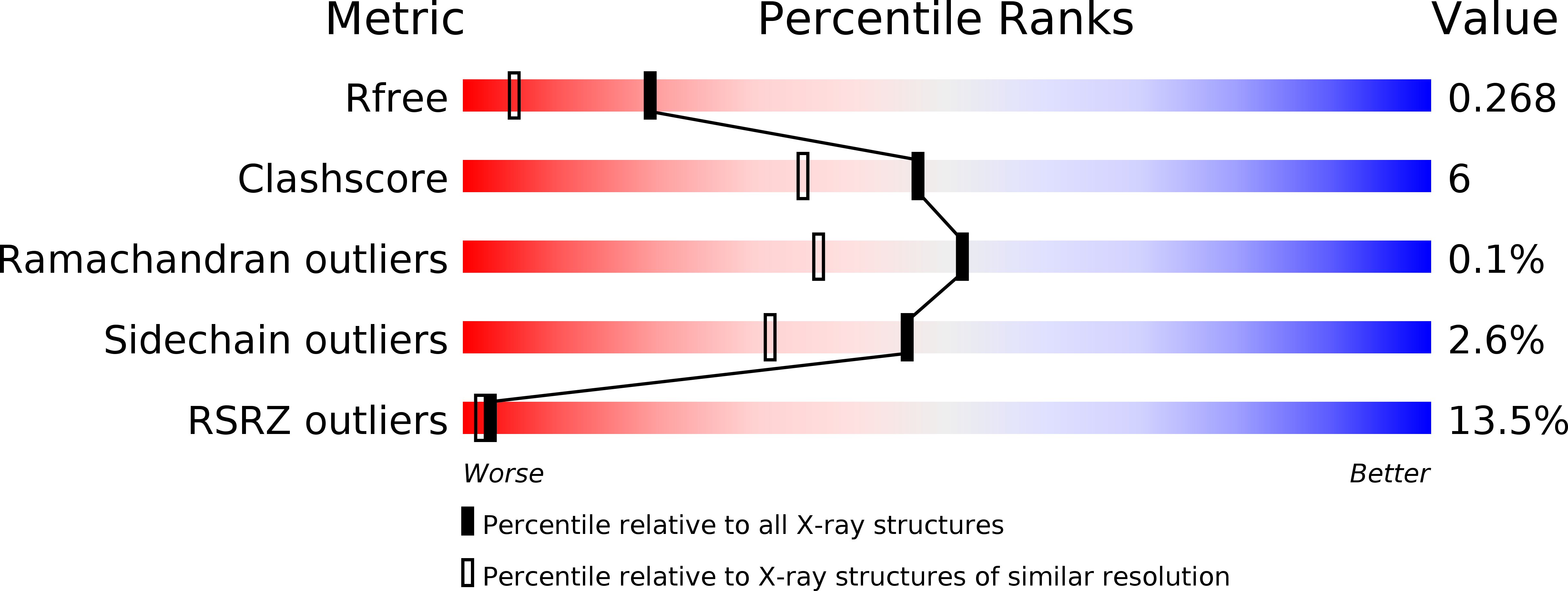
2H0Q image
Deposition Date 2006-05-15
Release Date 2007-11-06
Last Version Date 2024-02-14
Entry Detail
PDB ID:
2H0Q
Title:
Crystal Structure of the PGM domain of the Suppressor of T-Cell receptor (Sts-1)
Biological Source:
Source Organism:
Mus musculus (Taxon ID: 10090)
Host Organism:
Method Details:
Experimental Method:
Resolution:
1.82 Å
R-Value Free:
0.23
R-Value Work:
0.19
R-Value Observed:
0.19
Space Group:
C 1 2 1
Macromolecular Entities
Polymer Type:polypeptide(L)
Molecule:Suppressor of T-cell receptor signaling 1
Gene (Uniprot):Ubash3b
Chain IDs:A, B, C
Chain Length:261
Number of Molecules:3
Biological Source:Mus musculus
Primary Citation
A phosphatase activity of Sts-1 contributes to the suppression of TCR signaling.
Mol.Cell 27 486 497 (2007)
PMID: 17679096 DOI: 10.1016/j.molcel.2007.06.015

Abstact

Precise signaling by the T cell receptor (TCR) is crucial for a proper immune response. To ensure that T cells respond appropriately to antigenic stimuli, TCR signaling pathways are subject to multiple levels of regulation. Sts-1 negatively regulates signaling pathways downstream of the TCR by an unknown mechanism(s). Here, we demonstrate that Sts-1 is a phosphatase that can target the tyrosine kinase Zap-70 among other proteins. The X-ray structure of the Sts-1 C terminus reveals that it has homology to members of the phosphoglycerate mutase/acid phosphatase (PGM/AcP) family of enzymes, with residues known to be important for PGM/AcP catalytic activity conserved in nature and position in Sts-1. Point mutations that impair Sts-1 phosphatase activity in vitro also impair the ability of Sts-1 to regulate TCR signaling in T cells. These observations reveal a PGM/AcP-like enzyme activity involved in the control of antigen receptor signaling.

Legend

Protein

Chemical

Disease

Primary Citation of related structures