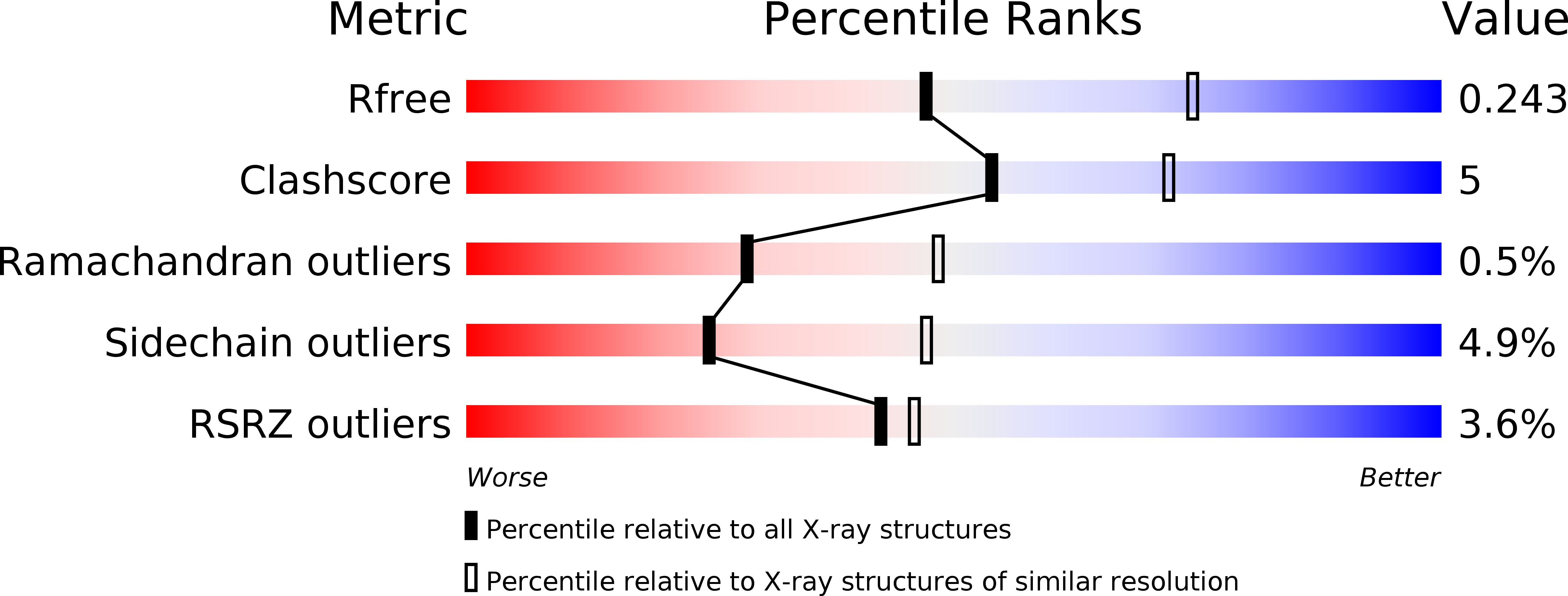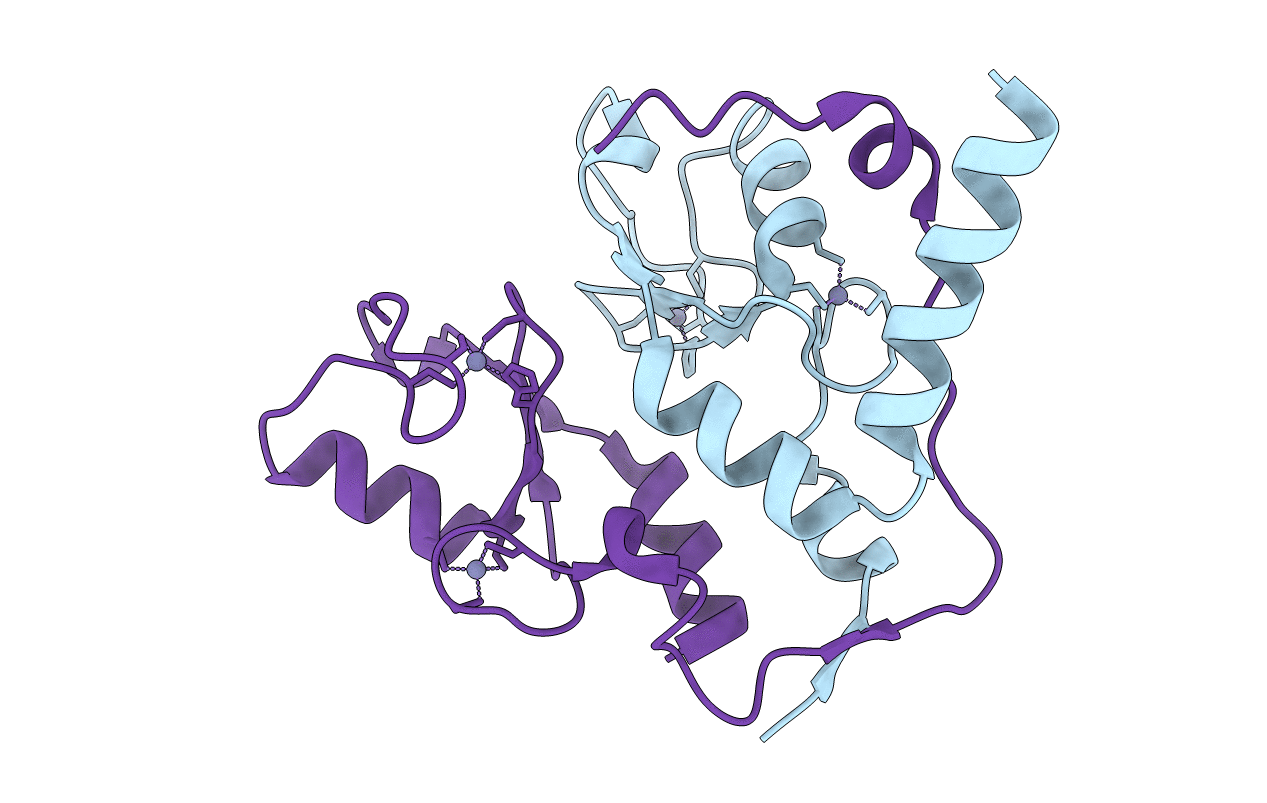
2H0D image
Deposition Date 2006-05-14
Release Date 2006-05-23
Last Version Date 2024-02-14
Entry Detail
PDB ID:
2H0D
Title:
Structure of a Bmi-1-Ring1B Polycomb group ubiquitin ligase complex
Biological Source:
Source Organism:
Homo sapiens (Taxon ID: 9606)
Method Details:
Experimental Method:
Resolution:
2.50 Å
R-Value Free:
0.24
R-Value Work:
0.20
R-Value Observed:
0.21
Space Group:
P 63
Macromolecular Entities
Polymer Type:polypeptide(L)
Molecule:B lymphoma Mo-MLV insertion region
Chain IDs:A
Chain Length:97
Number of Molecules:1
Biological Source:Homo sapiens
Polymer Type:polypeptide(L)
Molecule:Ubiquitin ligase protein RING2
Gene (Uniprot):RNF2
Chain IDs:B
Chain Length:100
Number of Molecules:1
Biological Source:Homo sapiens
Ligand Molecules
Primary Citation
Structure of a Bmi-1-Ring1B Polycomb Group Ubiquitin Ligase Complex.
J.Biol.Chem. 281 20643 20649 (2006)
PMID: 16714294 DOI: 10.1074/jbc.M602461200

Abstact

Polycomb group proteins Bmi-1 and Ring1B are core subunits of the PRC1 complex, which plays important roles in the regulation of Hox gene expression, X-chromosome inactivation, tumorigenesis, and stem cell self-renewal. The RING finger protein Ring1B is an E3 ligase that participates in the ubiquitination of lysine 119 of histone H2A, and the binding of Bmi-1 stimulates the E3 ligase activity. We have mapped the regions of Bmi-1 and Ring1B required for efficient ubiquitin transfer and determined a 2.5-A structure of the Bmi-1-Ring1B core domain complex. The structure reveals that Ring1B "hugs" Bmi-1 through extensive RING domain contacts and its N-terminal tail wraps around Bmi-1. The two regions of interaction have a synergistic effect on the E3 ligase activity. Our analyses suggest a model where the Bmi-1-Ring1B complex stabilizes the interaction between the E2 enzyme and the nucleosomal substrate to allow efficient ubiquitin transfer.

Legend

Protein

Chemical

Disease

Primary Citation of related structures