2GZ5 image
Deposition Date 2006-05-10
Release Date 2006-05-23
Last Version Date 2024-10-30
Entry Detail
PDB ID:
2GZ5
Keywords:
Title:
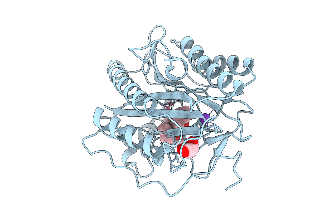
Human Type 1 methionine aminopeptidase in complex with ovalicin at 1.1 Ang
Biological Source:
Source Organism:
Homo sapiens (Taxon ID: 9606)
Method Details:
Experimental Method:
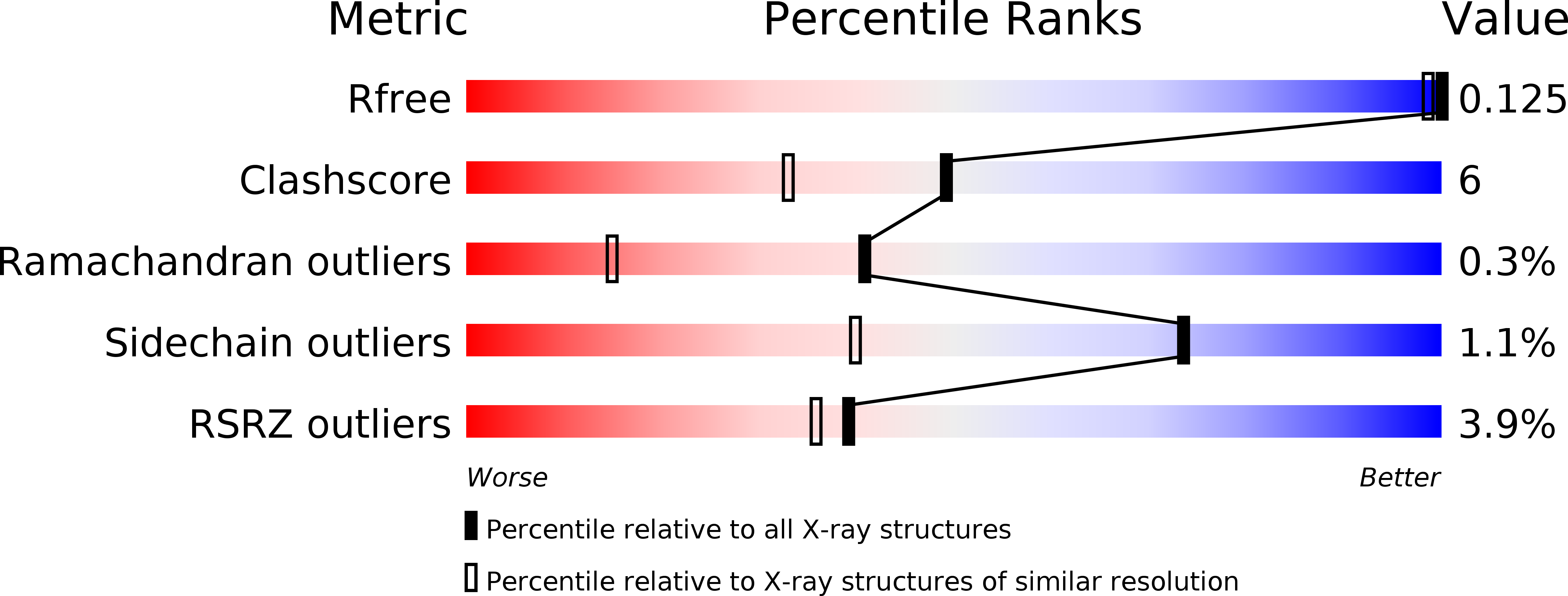
Resolution:
1.10 Å
R-Value Free:
0.14
R-Value Work:
0.11
R-Value Observed:
0.13
Space Group:
P 1 21 1
Macromolecular Entities
Polymer Type:polypeptide(L)
Molecule:Methionine aminopeptidase 1
Gene (Uniprot):METAP1
Chain IDs:A
Chain Length:329
Number of Molecules:1
Biological Source:Homo sapiens
Primary Citation
Structure of the angiogenesis inhibitor ovalicin bound to its noncognate target, human Type 1 methionine aminopeptidase.
Protein Sci. 15 1842 1848 (2006)
PMID: 16823043 DOI: 10.1110/ps.062278006

Abstact

Methionine aminopeptidases (MetAPs) remove the initiator methionine during protein biosynthesis. They exist in two isoforms, MetAP1 and MetAP2. The anti-angiogenic compound fumagillin binds tightly to the Type 2 MetAPs but only weakly to Type 1. High-affinity complexes of fumagillin and its relative ovalicin with Type 2 human MetAP have been reported. Here we describe the crystallographic structure of the low-affinity complex between ovalicin and Type 1 human MetAP at 1.1 A resolution. This provides the first opportunity to compare the structures of ovalicin or fumagillin bound to a Type 1 and a Type 2 MetAP. For both Type 1 and Type 2 human MetAPs the inhibitor makes a covalent adduct with a corresponding histidine. At the same time there are significant differences in the alignment of the inhibitors within the respective active sites. It has been argued that the lower affinity of ovalicin and fumagillin for the Type 1 MetAPs is due to the smaller size of their active sites relative to the Type 2 enzymes. Comparison with the uncomplexed structure of human Type 1 MetAP indicates that there is some truth to this. Several active site residues have to move "outward" by 0.5 Angstroms or so to accommodate the inhibitor. Other residues move "inward." There are, however, other factors that come into play. In particular, the side chain of His310 rotates by 134 degrees into a different position where (together with Glu128 and Tyr195) it coordinates a metal ion not seen at this site in the native enzyme.

Legend

Protein

Chemical

Disease

Primary Citation of related structures