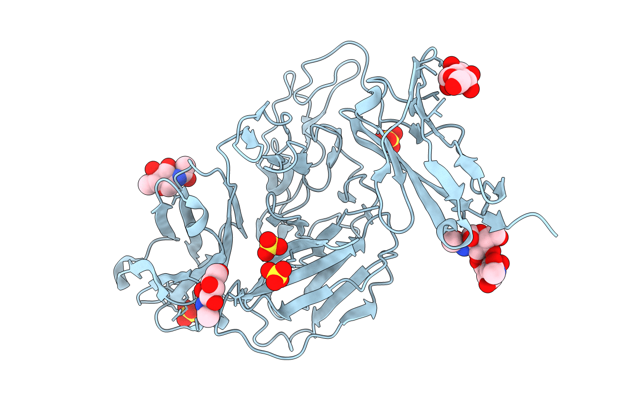
2GY5 image
Entry Detail
PDB ID:
2GY5
Keywords:
Title:
Tie2 Ligand-Binding Domain Crystal Structure
Biological Source:
Source Organism:
Host Organism:
PDB Version:
Deposition Date:
2006-05-09
Release Date:
2006-06-06
Method Details:
Experimental Method:
Resolution:
2.90 Å
R-Value Free:
0.29
R-Value Work:
0.24
R-Value Observed:
0.25
Space Group:
P 41 21 2
Macromolecular Entities
Polymer Type:polypeptide(L)
Description:Angiopoietin-1 receptor
Chain IDs:A
Chain Length:423
Number of Molecules:1
Biological Source:Homo sapiens
Modified Residue
Compound ID Chain ID Parent Comp ID Details 2D Image
ASN A ASN GLYCOSYLATION SITE
Primary Citation
Crystal structures of the Tie2 receptor ectodomain and the angiopoietin-2-Tie2 complex.
Nat.Struct.Mol.Biol. 13 524 532 (2006)
PMID: 16732286 DOI: 10.1038/nsmb1101

Abstact

The Tie receptor tyrosine kinases and their angiopoietin (Ang) ligands play central roles in developmental and tumor-induced angiogenesis. Here we present the crystal structures of the Tie2 ligand-binding region alone and in complex with Ang2. In contrast to prediction, Tie2 contains not two but three immunoglobulin (Ig) domains, which fold together with the three epidermal growth factor domains into a compact, arrowhead-shaped structure. Ang2 binds at the tip of the arrowhead utilizing a lock-and-key mode of ligand recognition-unique for a receptor kinase-where two complementary surfaces interact with each other with no domain rearrangements and little conformational change in either molecule. Ang2-Tie2 recognition is similar to antibody-protein antigen recognition, including the location of the ligand-binding site within the Ig fold. Analysis of the structures and structure-based mutagenesis provide insight into the mechanism of receptor activation and support the hypothesis that all angiopoietins interact with Tie2 in a structurally similar manner.

Legend

Protein

Chemical

Disease

Primary Citation of related structures