2GWK image
Deposition Date 2006-05-04
Release Date 2006-08-29
Last Version Date 2025-03-26
Entry Detail
PDB ID:
2GWK
Title:
SpvB ADP-ribosylated actin: orthorhombic crystal form
Biological Source:
Source Organism:
Method Details:
Experimental Method:
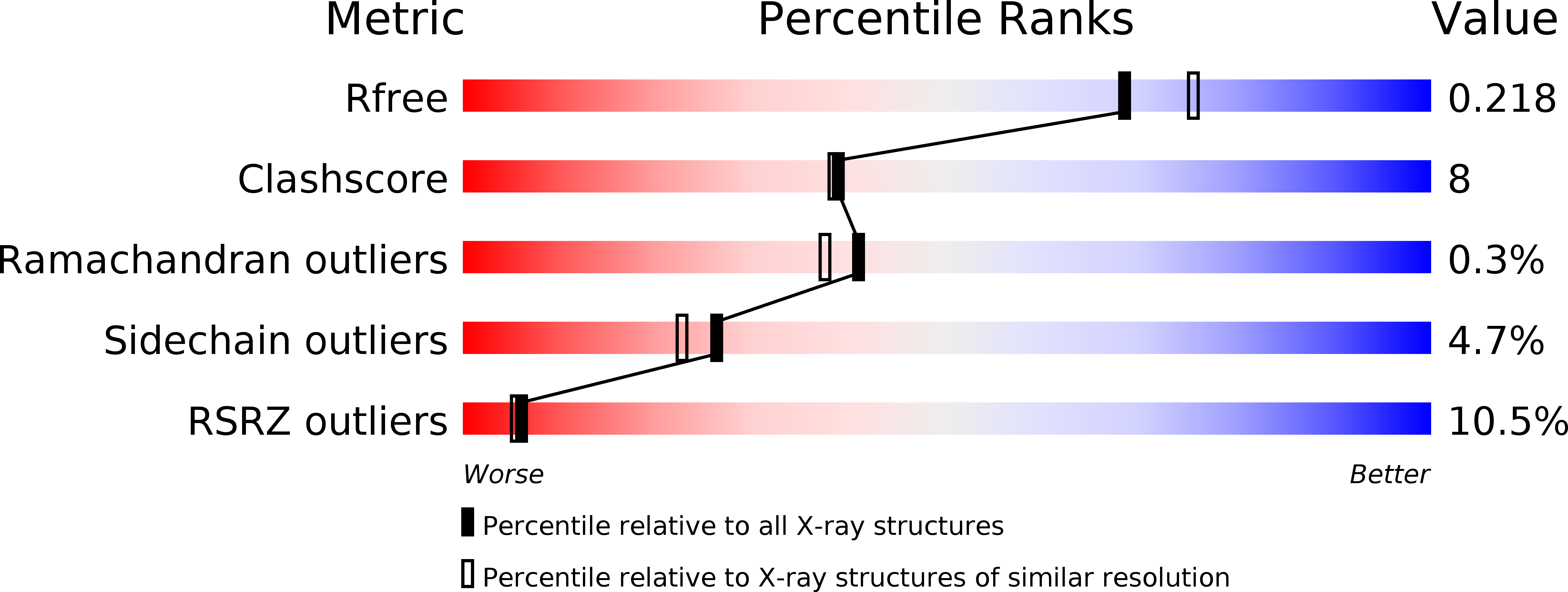
Resolution:
2.00 Å
R-Value Free:
0.21
R-Value Work:
0.17
R-Value Observed:
0.17
Space Group:
P 21 21 21
Macromolecular Entities
Polymer Type:polypeptide(L)
Molecule:Actin, alpha skeletal muscle
Gene (Uniprot):ACTA1
Chain IDs:A, B
Chain Length:371
Number of Molecules:2
Biological Source:Oryctolagus cuniculus
Modified Residue
Compound ID Chain ID Parent Comp ID Details 2D Image
HIC A HIS 4-METHYL-HISTIDINE
Primary Citation
A steric antagonism of actin polymerization by a salmonella virulence protein.
Structure 14 1219 1229 (2006)
PMID: 16905096 DOI: 10.1016/j.str.2006.05.022

Abstact

Salmonella spp. require the ADP-ribosyltransferase activity of the SpvB protein for intracellular growth and systemic virulence. SpvB covalently modifies actin, causing cytoskeletal disruption and apoptosis. We report here the crystal structure of the catalytic domain of SpvB, and we show by mass spectrometric analysis that SpvB modifies actin at Arg177, inhibiting its ATPase activity. We also describe two crystal structures of SpvB-modified, polymerization-deficient actin. These structures reveal that ADP-ribosylation does not lead to dramatic conformational changes in actin, suggesting a model in which this large family of toxins inhibits actin polymerization primarily through steric disruption of intrafilament contacts.

Legend

Protein

Chemical

Disease

Primary Citation of related structures