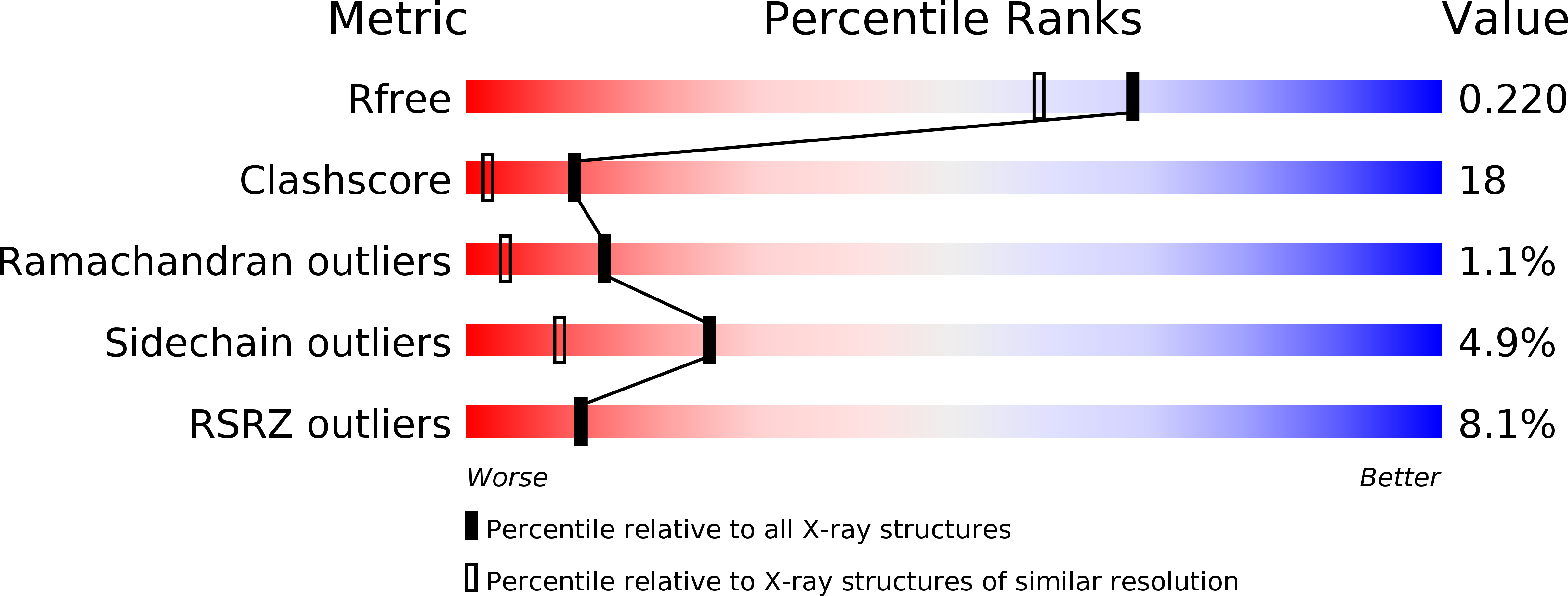
2GW8 image
Deposition Date 2006-05-04
Release Date 2006-06-13
Last Version Date 2023-08-30
Entry Detail
PDB ID:
2GW8
Title:
Structure of the PII signal transduction protein of Neisseria meningitidis at 1.85 resolution
Biological Source:
Source Organism:
Host Organism:
Method Details:
Experimental Method:
Resolution:
1.85 Å
R-Value Free:
0.21
R-Value Work:
0.18
R-Value Observed:
0.18
Space Group:
P 63
Macromolecular Entities
Structures with similar UniProt ID
Protein Blast
Polymer Type:polypeptide(L)
Molecule:PII signal transduction protein
Chain IDs:A
Chain Length:114
Number of Molecules:1
Biological Source:Neisseria meningitidis
Primary Citation
Structure of the P(II) signal transduction protein of Neisseria meningitidis at 1.85 A resolution.
Acta Crystallogr.,Sect.F 62 494 497 (2006)
PMID: 16754965 DOI: 10.1107/S1744309106015430

Abstact

The P(II) signal transduction proteins GlnB and GlnK are implicated in the regulation of nitrogen assimilation in Escherichia coli and other enteric bacteria. P(II)-like proteins are widely distributed in bacteria, archaea and plants. In contrast to other bacteria, Neisseria are limited to a single P(II) protein (NMB 1995), which shows a high level of sequence identity to GlnB and GlnK from Escherichia coli (73 and 62%, respectively). The structure of the P(II) protein from N. meningitidis (serotype B) has been solved by molecular replacement to a resolution of 1.85 A. Comparison of the structure with those of other P(II) proteins shows that the overall fold is tightly conserved across the whole population of related proteins, in particular the positions of the residues implicated in ATP binding. It is proposed that the Neisseria P(II) protein shares functions with GlnB/GlnK of enteric bacteria.

Legend

Protein

Chemical

Disease

Primary Citation of related structures