2GW3 image
Entry Detail
PDB ID:
2GW3
Title:
Crystal structure of stony coral fluorescent protein Kaede, green form
Biological Source:
Source Organism:
PDB Version:
Deposition Date:
2006-05-03
Release Date:
2007-05-08
Method Details:
Experimental Method:
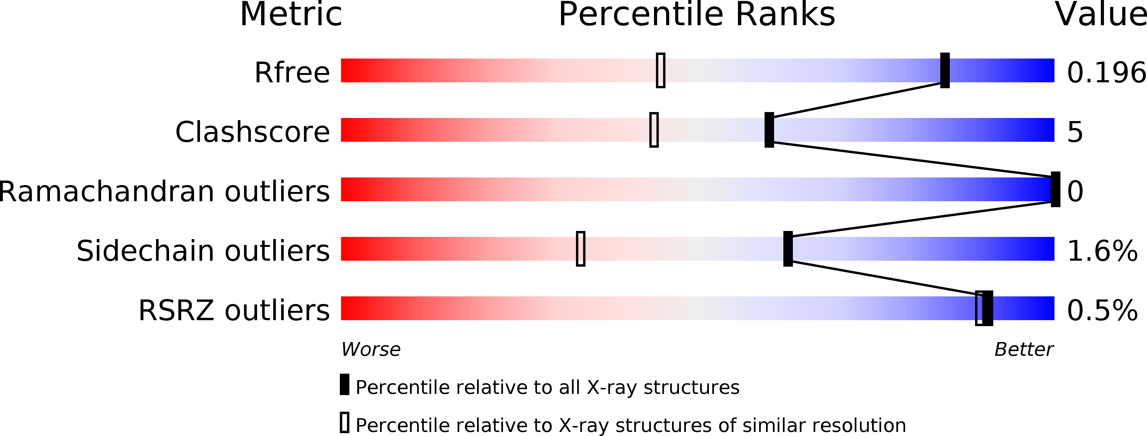
Resolution:
1.40 Å
R-Value Free:
0.21
R-Value Work:
0.19
Space Group:
C 1 2 1
Macromolecular Entities
Polymer Type:polypeptide(L)
Description:Kaede
Chain IDs:A, B
Chain Length:227
Number of Molecules:2
Biological Source:Trachyphyllia geoffroyi
Modified Residue
Compound ID Chain ID Parent Comp ID Details 2D Image
CR8 A HIS ?
Ligand Molecules
Primary Citation
Crystallographic evidence for water-assisted photo-induced peptide cleavage in the stony coral fluorescent protein Kaede.
J.Mol.Biol. 372 918 926 (2007)
PMID: 17692334 DOI: 10.1016/j.jmb.2007.06.037

Abstact

A coral fluorescent protein from Trachyphyllia geoffroyi, Kaede, possesses a tripeptide of His62-Tyr63-Gly64, which forms a chromophore with green fluorescence. This chromophore's fluorescence turns red following UV light irradiation. We have previously shown that such photoconversion is achieved by a formal beta-elimination reaction, which results in a cleavage of the peptide bond found between the amide nitrogen and the alpha-carbon at His62. However, the stereochemical arrangement of the chromophore and the precise structural basis for this reaction mechanism previously remained unknown. Here, we report the crystal structures of the green and red form of Kaede at 1.4 A and 1.6 A resolutions, respectively. Our structures depict the cleaved peptide bond in the red form. The chromophore conformations both in the green and red forms are similar, except a well-defined water molecule in the proximity of the His62 imidazole ring in the green form. We propose a molecular mechanism for green-to-red photoconversion, which is assisted by the water molecule.

Legend

Protein

Chemical

Disease

Primary Citation of related structures