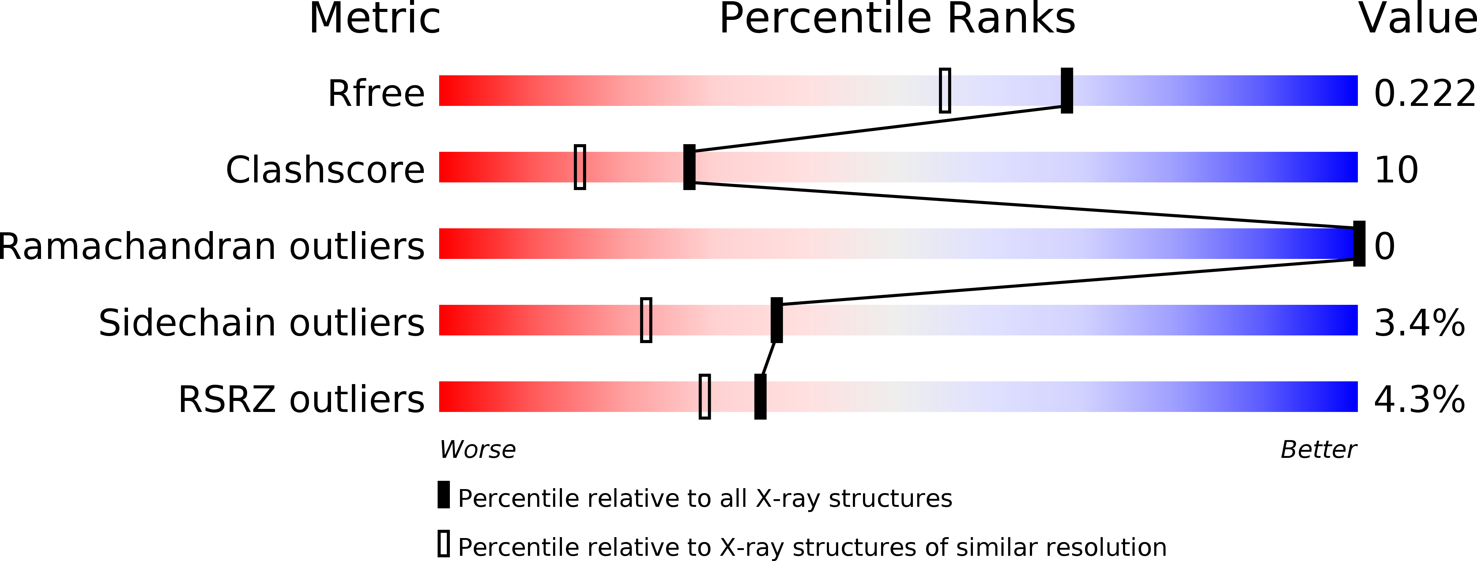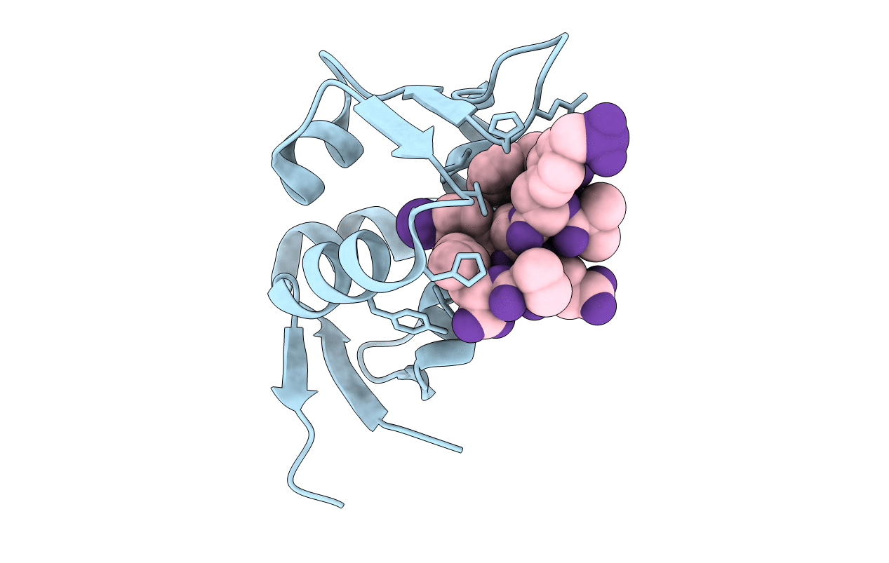
2GV2 image
Deposition Date 2006-05-02
Release Date 2006-09-05
Last Version Date 2023-11-15
Entry Detail
PDB ID:
2GV2
Keywords:
Title:
MDM2 in complex with an 8-mer p53 peptide analogue
Biological Source:
Source Organism:
Homo sapiens (Taxon ID: 9606)
(Taxon ID: )
Host Organism:
Method Details:
Experimental Method:
Resolution:
1.80 Å
R-Value Free:
0.21
R-Value Work:
0.19
R-Value Observed:
0.19
Space Group:
P 21 21 21
Macromolecular Entities
Polymer Type:polypeptide(L)
Molecule:E3 ubiquitin-protein ligase Mdm2
Gene (Uniprot):MDM2
Chain IDs:A
Chain Length:110
Number of Molecules:1
Biological Source:Homo sapiens
Polymer Type:polypeptide(L)
Molecule:8-MER P53 PEPTIDE ANALOGUE
Chain IDs:B
Chain Length:9
Number of Molecules:1
Biological Source:
Modified Residue
Compound ID Chain ID Parent Comp ID Details 2D Image
6CW B TRP 6-CHLORO-L-TRYPTOPHAN
AIB B ALA ALPHA-AMINOISOBUTYRIC ACID
PM3 B PHE ?
Primary Citation
Crystallographic Analysis of an 8-mer p53 Peptide Analogue Complexed with MDM2.
J.Am.Chem.Soc. 128 11000 11001 (2006)
PMID: 16925398 DOI: 10.1021/ja063102j

Abstact

The most potent inhibitor of the p53-MDM2 interaction reported to date is an 8-mer p53 peptide analogue (Novartis peptide), which contains 6-chlorotryptophane (Cl-Trp) and phosphonomethylphenylalanine (Pmp) as key residues for the enhanced activity. We report here a crystal structure of the co-complex between MDM2 and the Novartis peptide solved at 1.8 A resolution. The structural basis for the role of the two aromatic residues are delineated by comparing the present structure with crystal structures of the MDM2 co-complex bound to other inhibitors including the wt-p53 peptide itself.

Legend

Protein

Chemical

Disease

Primary Citation of related structures