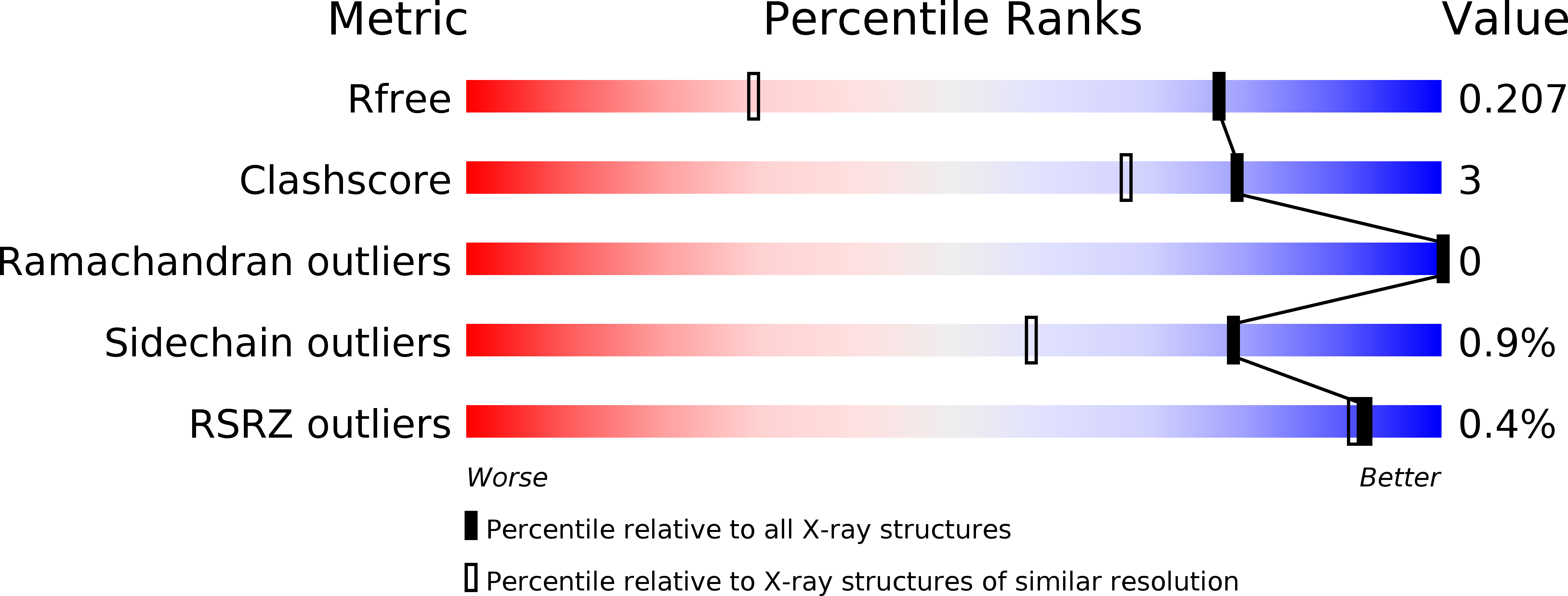
2GTE image
Deposition Date 2006-04-27
Release Date 2007-06-12
Last Version Date 2024-11-13
Entry Detail
PDB ID:
2GTE
Title:
Drosophila OBP LUSH bound to attractant pheromone 11-cis-vaccenyl acetate
Biological Source:
Source Organism:
Host Organism:
Method Details:
Experimental Method:
Resolution:
1.40 Å
R-Value Free:
0.20
R-Value Work:
0.17
R-Value Observed:
0.17
Space Group:
P 21 21 21
Macromolecular Entities
Polymer Type:polypeptide(L)
Molecule:General odorant-binding protein lush
Gene (Uniprot):lush
Chain IDs:A, B
Chain Length:124
Number of Molecules:2
Biological Source:Drosophila melanogaster
Primary Citation
Activation of pheromone-sensitive neurons is mediated by conformational activation of pheromone-binding protein
Cell(Cambridge,Mass.) 133 1255 1265 (2008)
PMID: 18585358 DOI: 10.1016/j.cell.2008.04.046

Abstact

Detection of volatile odorants by olfactory neurons is thought to result from direct activation of seven-transmembrane odorant receptors by odor molecules. Here, we show that detection of the Drosophila pheromone, 11-cis vaccenyl acetate (cVA), is instead mediated by pheromone-induced conformational shifts in the extracellular pheromone-binding protein, LUSH. We show that LUSH undergoes a pheromone-specific conformational change that triggers the firing of pheromone-sensitive neurons. Amino acid substitutions in LUSH that are predicted to reduce or enhance the conformational shift alter sensitivity to cVA as predicted in vivo. One substitution, LUSH(D118A), produces a dominant-active LUSH protein that stimulates T1 neurons through the neuronal receptor components Or67d and SNMP in the complete absence of pheromone. Structural analysis of LUSH(D118A) reveals that it closely resembles cVA-bound LUSH. Therefore, the pheromone-binding protein is an inactive, extracellular ligand converted by pheromone molecules into an activator of pheromone-sensitive neurons and reveals a distinct paradigm for detection of odorants.

Legend

Protein

Chemical

Disease

Primary Citation of related structures