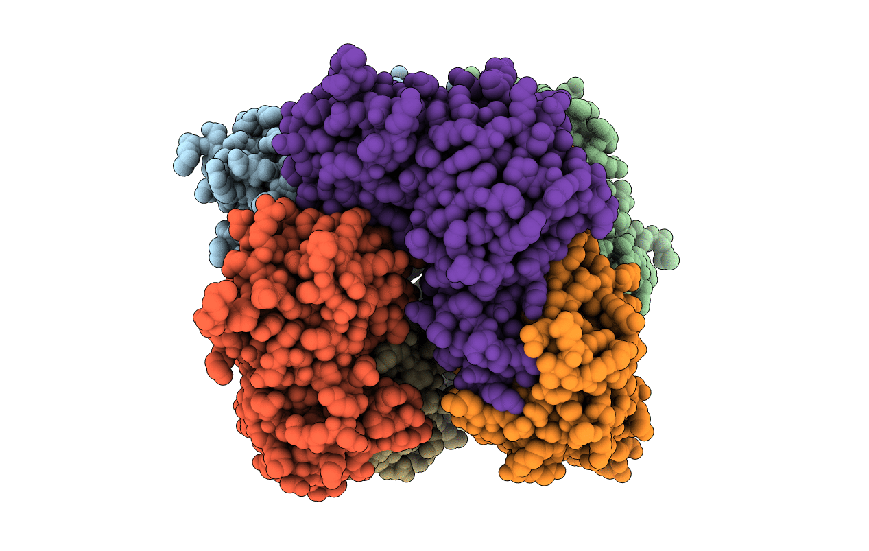
2GTD image
Deposition Date 2006-04-27
Release Date 2006-08-01
Last Version Date 2024-02-14
Entry Detail
PDB ID:
2GTD
Keywords:
Title:
Crystal Structure of a Type III Pantothenate Kinase: Insight into the Catalysis of an Essential Coenzyme A Biosynthetic Enzyme Universally Distributed in Bacteria
Biological Source:
Source Organism:
Method Details:
Experimental Method:
Resolution:
2.00 Å
R-Value Free:
0.24
R-Value Work:
0.18
R-Value Observed:
0.18
Space Group:
P 1 21 1
Macromolecular Entities
Polymer Type:polypeptide(L)
Molecule:Type III Pantothenate Kinase
Gene (Uniprot):coaX
Chain IDs:A, B, C, D, E, F
Chain Length:251
Number of Molecules:6
Biological Source:Thermotoga maritima
Primary Citation
Crystal Structure of a Type III Pantothenate Kinase: Insight into the Mechanism of an Essential Coenzyme A Biosynthetic Enzyme Universally Distributed in Bacteria.
J.Bacteriol. 188 5532 5540 (2006)
PMID: 16855243 DOI: 10.1128/JB.00469-06

Abstact

Pantothenate kinase (PanK) catalyzes the first step in the five-step universal pathway of coenzyme A (CoA) biosynthesis, a key transformation that generally also regulates the intracellular concentration of CoA through feedback inhibition. A novel PanK protein encoded by the gene coaX was recently identified that is distinct from the previously characterized type I PanK (exemplified by the Escherichia coli coaA-encoded PanK protein) and type II eukaryotic PanKs and is not inhibited by CoA or its thioesters. This type III PanK, or PanK-III, is widely distributed in the bacterial kingdom and accounts for the only known PanK in many pathogenic species, such as Helicobacter pylori, Bordetella pertussis, and Pseudomonas aeruginosa. Here we report the first crystal structure of a type III PanK, the enzyme from Thermotoga maritima (PanK(Tm)), solved at 2.0-A resolution. The structure of PanK(Tm) reveals that type III PanKs belong to the acetate and sugar kinase/heat shock protein 70/actin (ASKHA) protein superfamily and that they retain the highly conserved active site motifs common to all members of this superfamily. Comparative structural analysis of the PanK(Tm) active site configuration and mutagenesis of three highly conserved active site aspartates identify these residues as critical for PanK-III catalysis. Furthermore, the analysis also provides an explanation for the lack of CoA feedback inhibition by the enzyme. Since PanK-III adopts a different structural fold from that of the E. coli PanK -- which is a member of the "P-loop kinase"superfamily -- this finding represents yet another example of convergent evolution of the same biological function from a different protein ancestor.

Legend

Protein

Chemical

Disease

Primary Citation of related structures