2GS4 image
Deposition Date 2006-04-25
Release Date 2006-10-17
Last Version Date 2024-10-30
Entry Detail
PDB ID:
2GS4
Title:
The crystal structure of the E.coli stress protein YciF.
Biological Source:
Source Organism:
Escherichia coli (Taxon ID: 562)
Method Details:
Experimental Method:
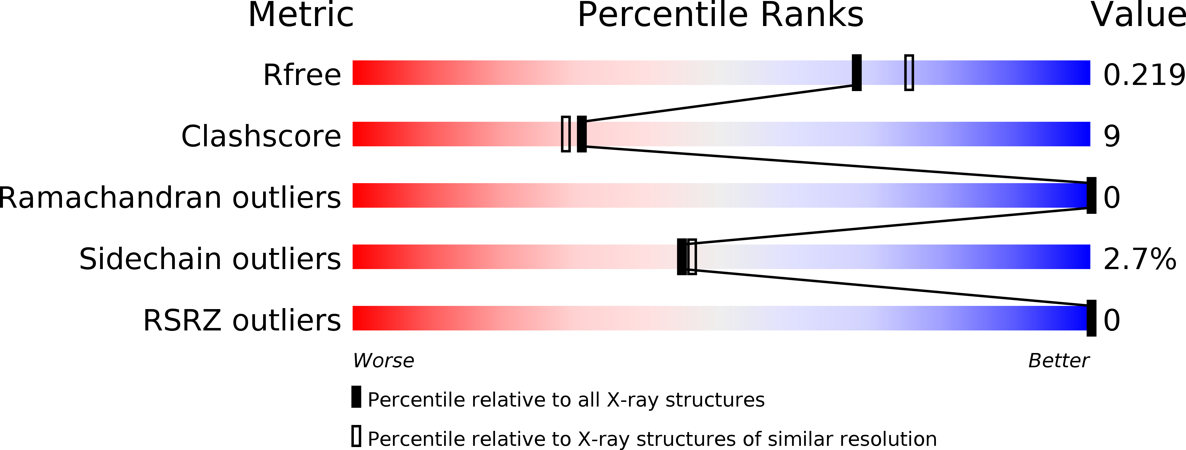
Resolution:
2.00 Å
R-Value Free:
0.22
R-Value Work:
0.18
R-Value Observed:
0.18
Space Group:
H 3
Macromolecular Entities
Polymer Type:polypeptide(L)
Molecule:Protein yciF
Gene (Uniprot):yciF
Chain IDs:A, B
Chain Length:166
Number of Molecules:2
Biological Source:Escherichia coli
Modified Residue
Compound ID Chain ID Parent Comp ID Details 2D Image
MSE A MET SELENOMETHIONINE
Primary Citation
The crystal structure of the E. coli stress protein YciF.
Protein Sci. 15 2605 2611 (2006)
PMID: 17001035 DOI: 10.1110/ps.062307706

Abstact

YciF is a protein that is up-regulated when bacteria experience stress conditions, and is highly conserved in a range of bacterial species. YciF has no known structure or biochemical function. To learn more about its potential molecular function and its role in the bacterial stress response, we solved the crystal structure of YciF at 2.0 Angstrom resolution by the multiple wavelength anomalous diffraction (MAD) technique. YciF is a dimer in solution, and forms a homodimer in the crystal asymmetric unit. The two monomers form a dimer with a molecular twofold axis, with a significant burial of solvent-accessible surface area. The protein is an all-alpha protein composed of five helices: a four-helix bundle, and a short additional helix at the dimer interface. The protein is structurally similar to portions of the diiron-containing proteins, rubrerythrin and the Bacillus anthracis Dlp-2.

Legend

Protein

Chemical

Disease

Primary Citation of related structures