
Deposition Date
2006-04-24
Release Date
2006-05-09
Last Version Date
2023-08-30
Entry Detail
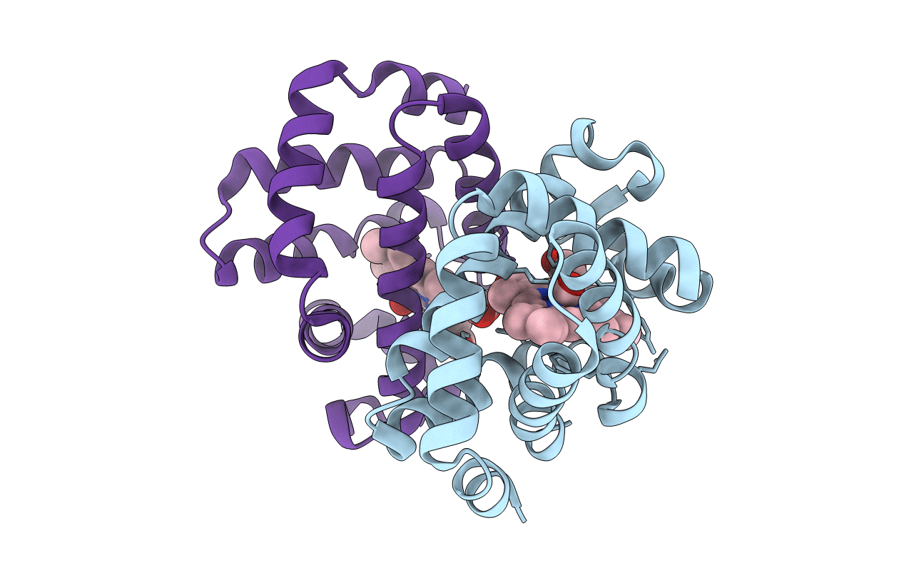
PDB ID:
2GRH
Keywords:
Title:
M37V mutant of Scapharca dimeric hemoglobin, with CO bound
Biological Source:
Source Organism(s):
Scapharca inaequivalvis (Taxon ID: 6561)
Expression System(s):
Method Details:
Experimental Method:
Resolution:
1.50 Å
R-Value Free:
0.19
R-Value Work:
0.18
R-Value Observed:
0.18
Space Group:
C 1 2 1
