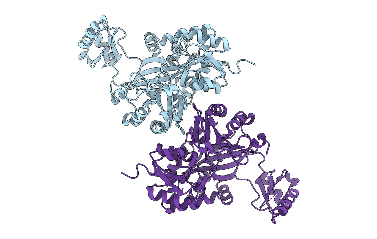
2GPS image
Deposition Date 2006-04-18
Release Date 2006-07-04
Last Version Date 2023-08-30
Entry Detail
PDB ID:
2GPS
Keywords:
Title:
Crystal Structure of the Biotin Carboxylase Subunit, E23R mutant, of Acetyl-CoA Carboxylase from Escherichia coli.
Biological Source:
Source Organism:
Escherichia coli (Taxon ID: 562)
Host Organism:
Method Details:
Experimental Method:
Resolution:
2.80 Å
R-Value Free:
0.25
R-Value Work:
0.20
R-Value Observed:
0.20
Space Group:
P 1 21 1
Macromolecular Entities
Polymer Type:polypeptide(L)
Molecule:Biotin carboxylase
Gene (Uniprot):accC
Mutations:E23R
Chain IDs:A, B
Chain Length:469
Number of Molecules:2
Biological Source:Escherichia coli
Primary Citation
Is dimerization required for the catalytic activity of bacterial biotin carboxylase?
Mol.Cell 22 807 818 (2006)
PMID: 16793549 DOI: 10.1016/j.molcel.2006.04.026

Abstact

Acetyl-coenzyme A carboxylases (ACCs) have crucial roles in fatty acid metabolism. The biotin carboxylase (BC) subunit of Escherichia coli ACC is believed to be active only as a dimer, although the crystal structure shows that the active site of each monomer is 25 A from the dimer interface. We report here biochemical, biophysical, and structural characterizations of BC carrying single-site mutations in the dimer interface. Our studies demonstrate that two of the mutants, R19E and E23R, are monomeric in solution but have only a 3-fold loss in catalytic activity. The crystal structures of the E23R and F363A mutants show that they can still form the correct dimer at high concentrations. Our data suggest that dimerization is not an absolute requirement for the catalytic activity of the E. coli BC subunit, and we propose a new model for the molecular mechanism of action for BC in multisubunit and multidomain ACCs.

Legend

Protein

Chemical

Disease

Primary Citation of related structures