2GOX image
Deposition Date 2006-04-14
Release Date 2007-03-20
Last Version Date 2024-11-06
Entry Detail
PDB ID:
2GOX
Title:
Crystal structure of Efb-C / C3d Complex
Biological Source:
Source Organism:
Method Details:
Experimental Method:
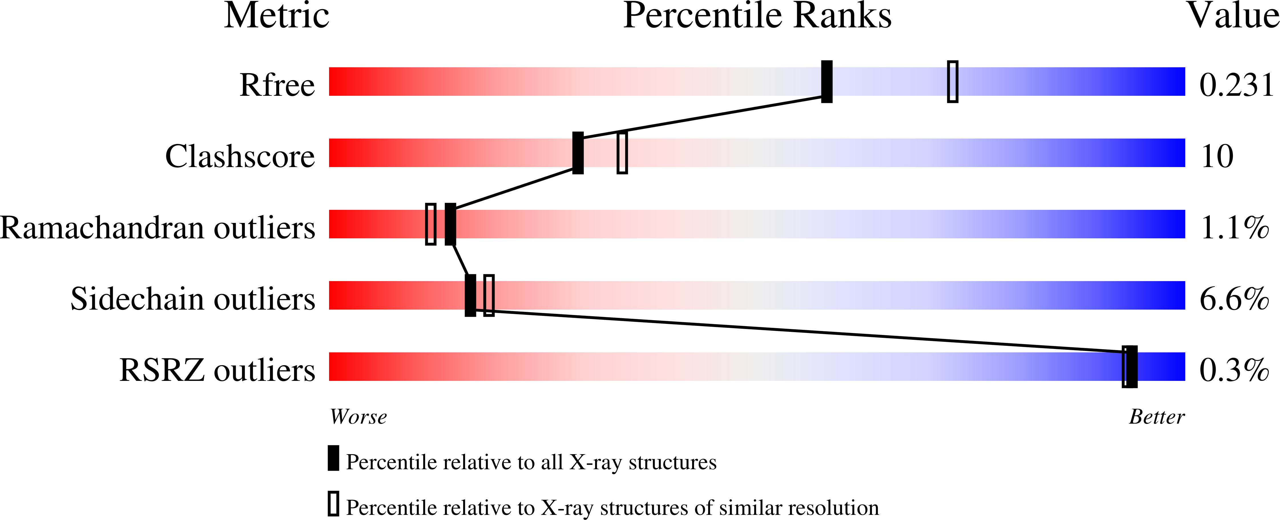
Resolution:
2.20 Å
R-Value Free:
0.23
R-Value Work:
0.18
Space Group:
P 41
Macromolecular Entities
Structures with similar UniProt ID
Protein Blast
Polymer Type:polypeptide(L)
Molecule:Complement C3
Gene (Uniprot):C3
Mutagens:C1010A
Chain IDs:A, C
Chain Length:297
Number of Molecules:2
Biological Source:Homo sapiens
Structures with similar UniProt ID
Protein Blast
Polymer Type:polypeptide(L)
Molecule:Fibrinogen-binding protein
Gene (Uniprot):fib
Chain IDs:B, D
Chain Length:65
Number of Molecules:2
Biological Source:Staphylococcus aureus subsp. aureus Mu50
Primary Citation
A structural basis for complement inhibition by Staphylococcus aureus.
Nat.Immunol. 8 430 437 (2007)
PMID: 17351618 DOI: 10.1038/ni1450

Abstact

To provide insight into bacterial suppression of complement-mediated immunity, we present here structures of a bacterial complement inhibitory protein, both free and bound to its complement target. The 1.25-A structure of the complement component C3-inhibitory domain of Staphylococcus aureus extracellular fibrinogen-binding protein (Efb-C) demonstrated a helical motif involved in complement regulation, whereas the 2.2-A structure of Efb-C bound to the C3d domain of human C3 allowed insight into the recognition of complement proteins by invading pathogens. Our structure-function studies provided evidence for a previously unrecognized mode of complement inhibition whereby Efb-C binds to native C3 and alters the solution conformation of C3 in a way that renders it unable to participate in successful 'downstream' activation of the complement response.

Legend

Protein

Chemical

Disease

Primary Citation of related structures