2GO4 image
Deposition Date 2006-04-12
Release Date 2006-07-04
Last Version Date 2023-08-30
Entry Detail
PDB ID:
2GO4
Keywords:
Title:
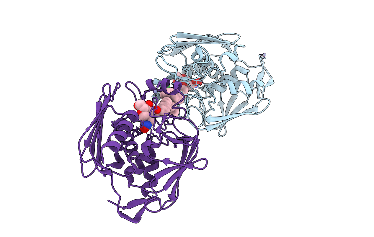
Crystal structure of Aquifex aeolicus LpxC complexed with TU-514
Biological Source:
Source Organism:
Host Organism:
Method Details:
Experimental Method:
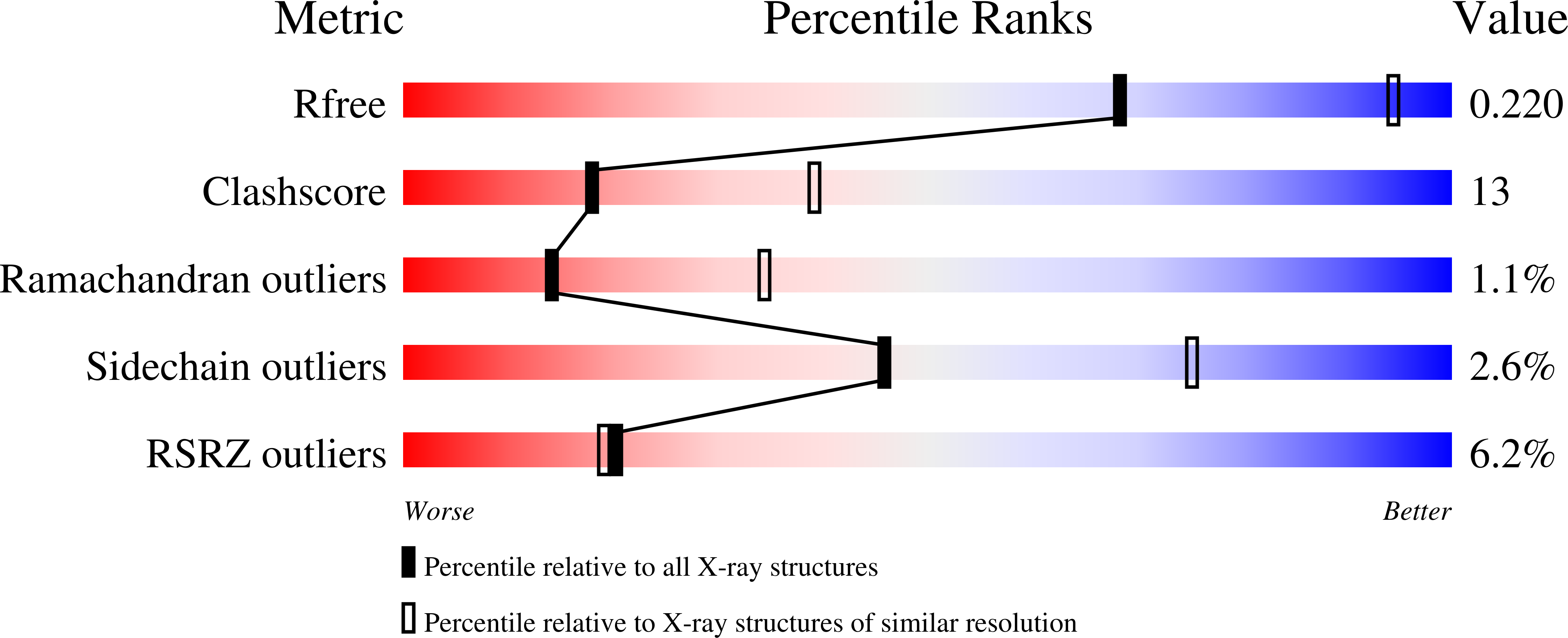
Resolution:
2.70 Å
R-Value Free:
0.24
R-Value Work:
0.20
Space Group:
P 61
Macromolecular Entities
Structures with similar UniProt ID
Protein Blast
Polymer Type:polypeptide(L)
Molecule:UDP-3-O-[3-hydroxymyristoyl] N-acetylglucosamine deacetylase
Gene (Uniprot):lpxC
Mutagens:C193A
Chain IDs:A, B
Chain Length:267
Number of Molecules:2
Biological Source:Aquifex aeolicus
Primary Citation
Mechanistic Inferences from the Binding of Ligands to LpxC, a Metal-Dependent Deacetylase
Biochemistry 45 7940 7948 (2006)
PMID: 16800620 DOI: 10.1021/bi060823m

Abstact

The metal-dependent deacetylase LpxC catalyzes the first committed step of lipid A biosynthesis in Gram-negative bacteria. Accordingly, LpxC is an attractive target for the development of inhibitors that may serve as potential new antibiotics for the treatment of Gram-negative bacterial infections. Here, we report the 2.7 A resolution X-ray crystal structure of LpxC complexed with the substrate analogue inhibitor TU-514 and the 2.0 A resolution structure of LpxC complexed with imidazole. The X-ray crystal structure of LpxC complexed with TU-514 allows for a detailed examination of the coordination geometry of the catalytic zinc ion and other enzyme-inhibitor interactions in the active site. The hydroxamate group of TU-514 forms a bidentate chelate complex with the zinc ion and makes hydrogen bond interactions with conserved active site residues E78, H265, and T191. The inhibitor C-4 hydroxyl group makes direct hydrogen bond interactions with E197 and H58. Finally, the C-3 myristate moiety of the inhibitor binds in the hydrophobic tunnel of the active site. These intermolecular interactions provide a foundation for understanding structural aspects of enzyme-substrate and enzyme-inhibitor affinity. Comparison of the TU-514 complex with cacodylate and imidazole complexes suggests a possible substrate diphosphate binding site and highlights residues that may stabilize the tetrahedral intermediate and its flanking transition states in catalysis. Evidence of a catalytic zinc ion in the native zinc enzyme coordinated by H79, H238, D242, and two water molecules with square pyramidal geometry is also presented. These results suggest that the native state of this metallohydrolase may contain a pentacoordinate zinc ion, which contrasts with the native states of archetypical zinc hydrolases such as thermolysin and carboxypeptidase A.

Legend

Protein

Chemical

Disease

Primary Citation of related structures