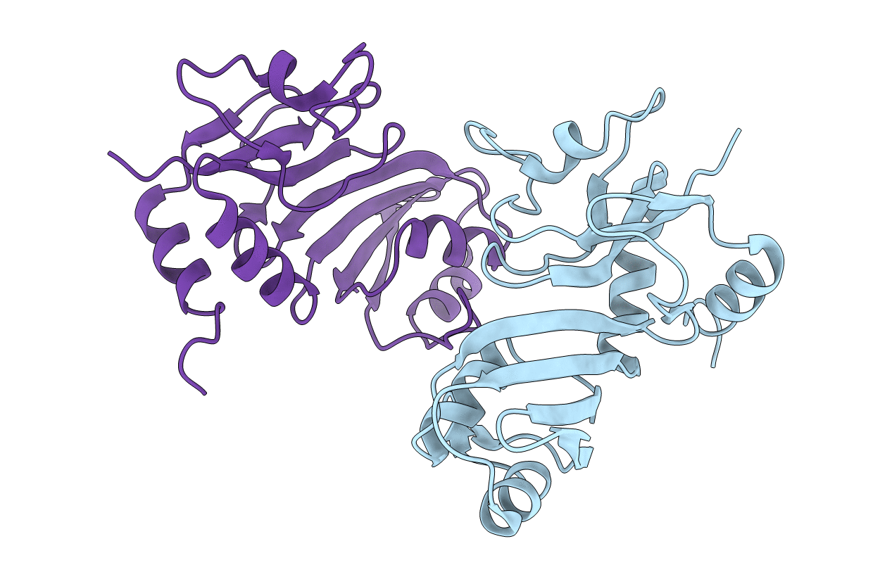
2GML image
Deposition Date 2006-04-06
Release Date 2006-07-18
Last Version Date 2024-11-13
Entry Detail
PDB ID:
2GML
Keywords:
Title:
Crystal Structure of Catalytic Domain of E.coli RluF
Biological Source:
Source Organism:
Escherichia coli (Taxon ID: 562)
Method Details:
Experimental Method:
Resolution:
2.60 Å
R-Value Free:
0.28
R-Value Work:
0.22
Space Group:
P 43 21 2
Macromolecular Entities
Structures with similar UniProt ID
Protein Blast
Polymer Type:polypeptide(L)
Molecule:Ribosomal large subunit pseudouridine synthase F
Gene (Uniprot):rluF
Chain IDs:A, B
Chain Length:237
Number of Molecules:2
Biological Source:Escherichia coli
Modified Residue
Compound ID Chain ID Parent Comp ID Details 2D Image
MSE A MET SELENOMETHIONINE
Primary Citation
Domain Organization and Crystal Structure of the Catalytic Domain of E.coli RluF, a Pseudouridine Synthase that Acts on 23S rRNA
J.Mol.Biol. 359 998 1009 (2006)
PMID: 16712869 DOI: 10.1016/j.jmb.2006.04.019

Abstact

Pseudouridine synthases catalyze the isomerization of uridine to pseudouridine (Psi) in rRNA and tRNA. The pseudouridine synthase RluF from Escherichia coli (E.C. 4.2.1.70) modifies U2604 in 23S rRNA, and belongs to a large family of pseudouridine synthases present in all kingdoms of life. Here we report the domain architecture and crystal structure of the catalytic domain of E.coli RluF at 2.6A resolution. Limited proteolysis, mass spectrometry and N-terminal sequencing indicate that RluF has a distinct domain architecture, with the catalytic domain flanked at the N and C termini by additional domains connected to it by flexible linkers. The structure of the catalytic domain of RluF is similar to those of RsuA and TruB. RluF is a member of the RsuA sequence family of Psi-synthases, along with RluB and RluE. Structural comparison of RluF with its closest structural homologues, RsuA and TruB, suggests possible functional roles for the N-terminal and C-terminal domains of RluF.

Legend

Protein

Chemical

Disease

Primary Citation of related structures