Deposition Date
2006-04-05
Release Date
2006-08-29
Last Version Date
2024-03-13
Entry Detail
PDB ID:
2GLX
Keywords:
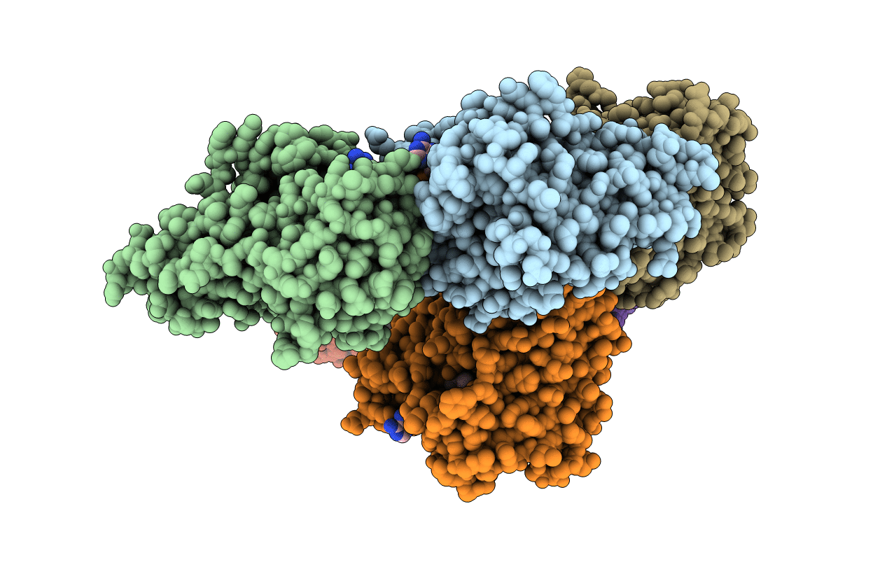
Title:
Crystal Structure Analysis of bacterial 1,5-AF Reductase
Biological Source:
Source Organism(s):
Ensifer adhaerens (Taxon ID: 106592)
Expression System(s):
Method Details:
Experimental Method:
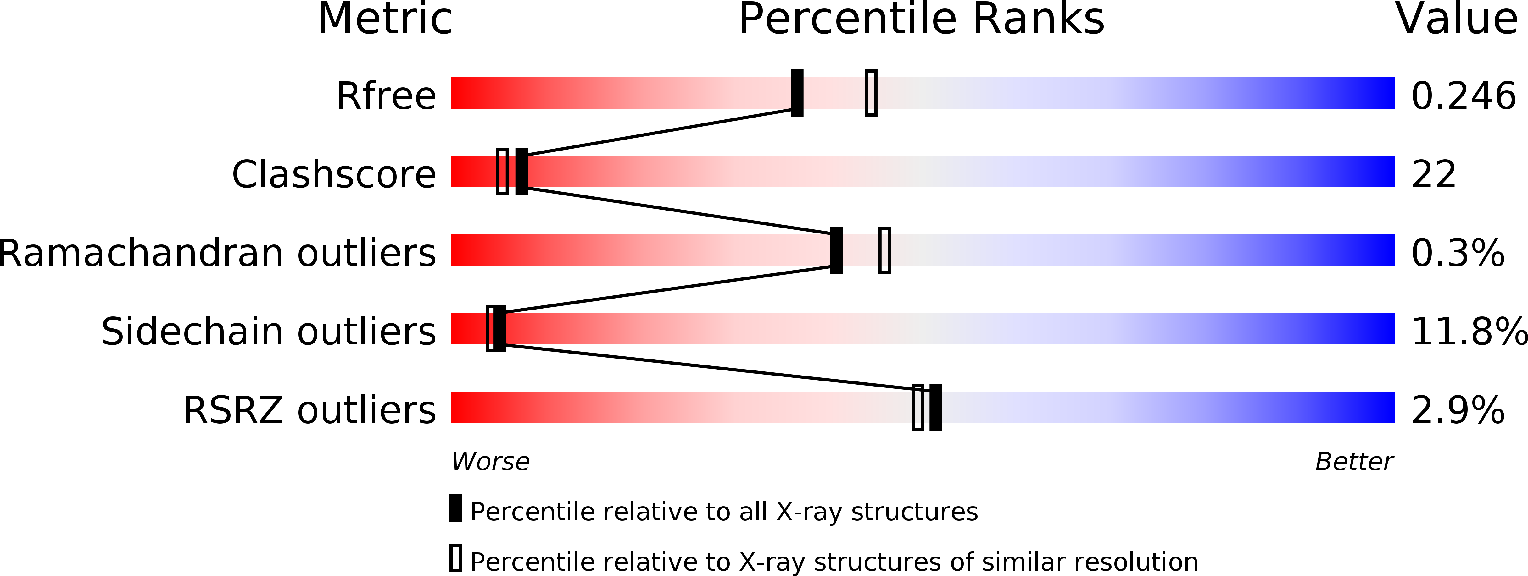
Resolution:
2.20 Å
R-Value Free:
0.25
R-Value Work:
0.19
R-Value Observed:
0.19
Space Group:
P 1 21 1