2GIX image
Deposition Date 2006-03-29
Release Date 2006-07-25
Last Version Date 2024-04-03
Entry Detail
PDB ID:
2GIX
Keywords:
Title:
Cytoplasmic Domain Structure of Kir2.1 containing Andersen's Mutation R218Q and Rescue Mutation T309K
Biological Source:
Source Organism:
Mus musculus (Taxon ID: 10090)
Host Organism:
Method Details:
Experimental Method:
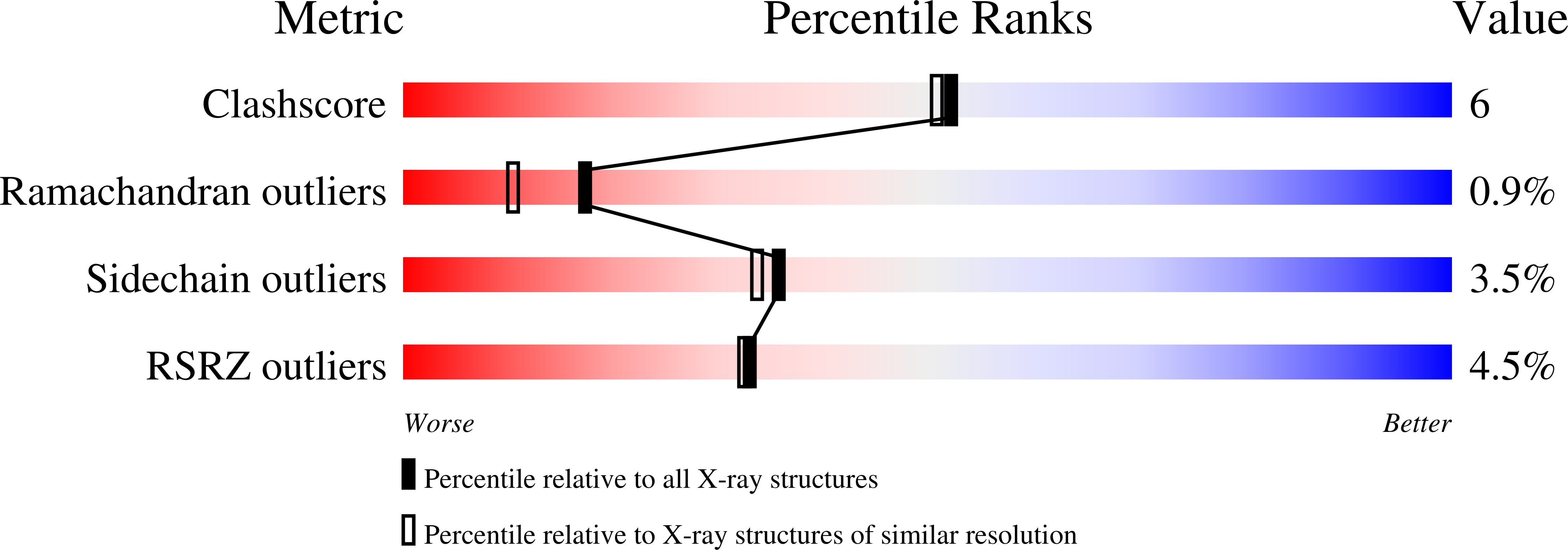
Resolution:
2.02 Å
R-Value Free:
0.22
R-Value Work:
0.17
R-Value Observed:
0.19
Space Group:
C 1 2 1
Macromolecular Entities
Structures with similar UniProt ID
Protein Blast
Polymer Type:polypeptide(L)
Molecule:Inward rectifier potassium channel 2
Gene (Uniprot):Kcnj2
Mutagens:R218Q, T309K
Chain IDs:A, B, C, D
Chain Length:208
Number of Molecules:4
Biological Source:Mus musculus
Primary Citation
Andersen's Syndrome Mutation Effects on the Structure and Assembly of the Cytoplasmic Domains of Kir2.1.
Biochemistry 45 8599 8606 (2006)
PMID: 16834334 DOI: 10.1021/bi060653d

Abstact

Kir2.1 channels play a key role in maintaining the correct resting potential in eukaryotic cells. Recently, specific amino acid mutations in the Kir2.1 inwardly rectifying potassium channel have been found to cause Andersen's Syndrome in humans. Here, we have characterized individual Andersen's Syndrome mutants R218Q, G300V, E303K, and delta314-315 and have found multiple effects on the ability of the cytoplasmic domains in Kir2.1 channels to form proper tetrameric assemblies. For the R218Q mutation, we identified a second site mutation (T309K) that restored tetrameric assembly but not function. We successfully crystallized and solved the structure (at 2.0 A) of the N- and C-terminal cytoplasmic domains of Kir2.1-R218Q/T309K(S). This new structure revealed multiple conformations of the G-loop and CD loop, providing an explanation for channels that assemble but do not conduct ions. Interestingly, Glu303 forms both intra- and intersubunit salt bridges, depending on the conformation of the G-loop, suggesting that the E303K mutant stabilizes both closed and open G-loop conformations. In the Kir2.1-R218Q/T309K(S) structure, we discovered that the DE loop forms a hydrophobic pocket that binds 2-methyl-2,4-pentanediol, which is located near the putative G(betagamma)-activation site of Kir3 channels. Finally, we observed a potassium ion bound to the cytoplasmic domain for this class of K+ channels.

Legend

Protein

Chemical

Disease

Primary Citation of related structures
Feedback Form
Name
Email
Institute
Feedback