Deposition Date
2006-03-29
Release Date
2006-04-11
Last Version Date
2023-08-30
Entry Detail
PDB ID:
2GIM
Keywords:
Title:
1.6 Angstrom structure of plastocyanin from Anabaena variabilis
Biological Source:
Source Organism(s):
Anabaena variabilis (Taxon ID: 1172)
Expression System(s):
Method Details:
Experimental Method:
Resolution:
1.60 Å
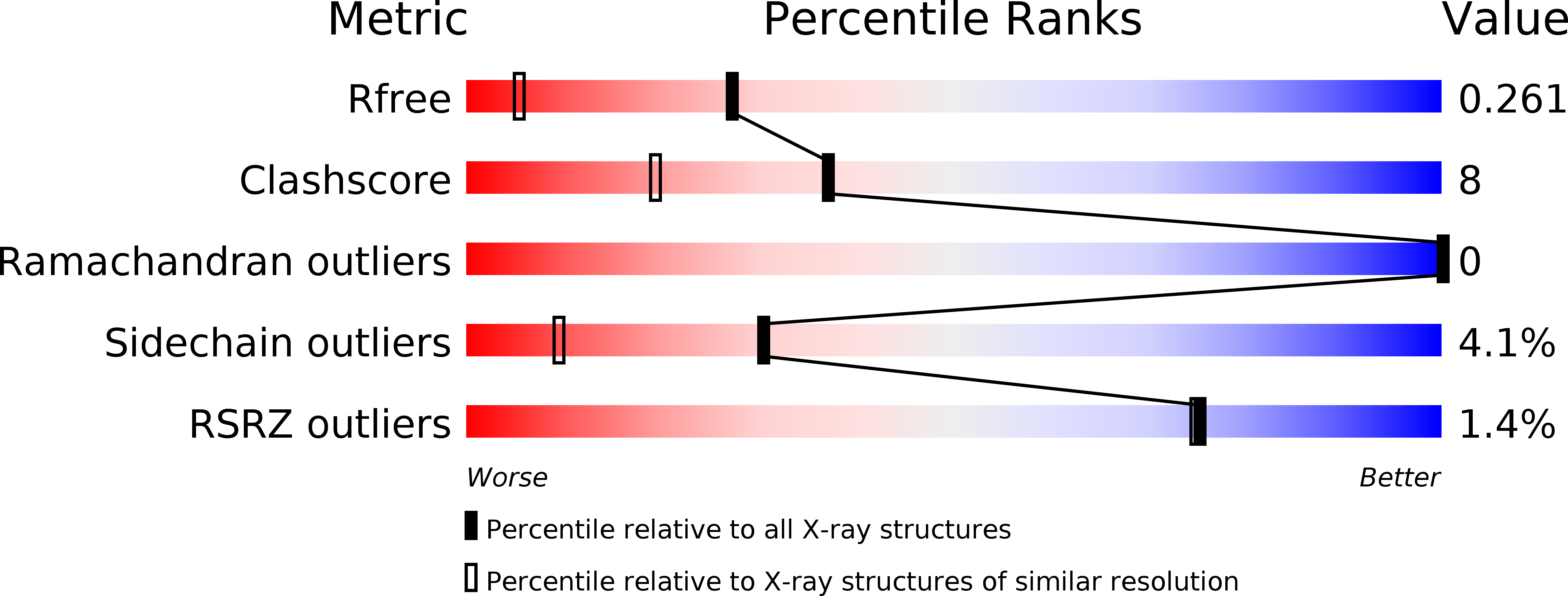
R-Value Free:
0.26
R-Value Work:
0.20
R-Value Observed:
0.21
Space Group:
P 21 21 21