2GFI image
Deposition Date 2006-03-22
Release Date 2007-03-27
Last Version Date 2024-10-30
Entry Detail
PDB ID:
2GFI
Keywords:
Title:
Crystal structure of the phytase from D. castellii at 2.3 A
Biological Source:
Source Organism:
Host Organism:
Method Details:
Experimental Method:
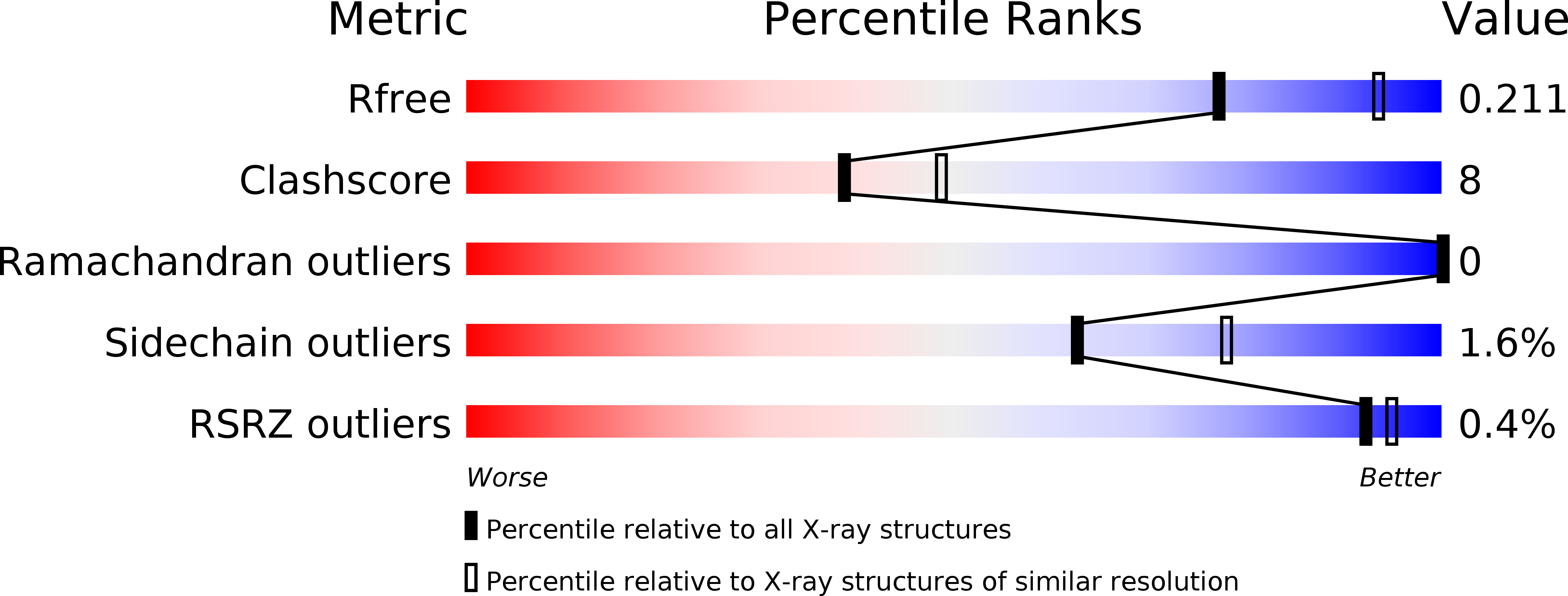
Resolution:
2.29 Å
R-Value Free:
0.21
R-Value Work:
0.15
Space Group:
P 65 2 2
Macromolecular Entities
Structures with similar UniProt ID
Protein Blast
Polymer Type:polypeptide(L)
Molecule:phytase
Gene (Uniprot):phyt
Chain IDs:A, B
Chain Length:458
Number of Molecules:2
Biological Source:Debaryomyces castellii
Modified Residue
Compound ID Chain ID Parent Comp ID Details 2D Image
ASN A ASN GLYCOSYLATION SITE
Ligand Molecules
Primary Citation
Structure of Debaryomyces castellii CBS 2923 phytase.
Acta Crystallogr.,Sect.F 65 321 326 (2009)
PMID: 19342770 DOI: 10.1107/S1744309109008653

Abstact

Phytate (myo-inositol hexakisphosphate) is the primary storage form of phosphate in seeds and legumes (Reddy et al., 1982). Phytases are phosphatases that hydrolyze phytate to less phosphorylated myo-inositol derivatives and inorganic phosphate. The crystal structure of phytase from Debaryomyces castellii has been determined at 2.3 A resolution. The crystals belonged to space group P6(5)22, with unit-cell parameters a = 121.65, c = 332.24 A. The structure was solved by molecular replacement and refined to a final R factor of 15.7% (R(free) = 20.9%). The final model consists of a dimer (with two monomers of 458 residues), five NAG molecules and 628 water molecules.

Legend

Protein

Chemical

Disease

Primary Citation of related structures