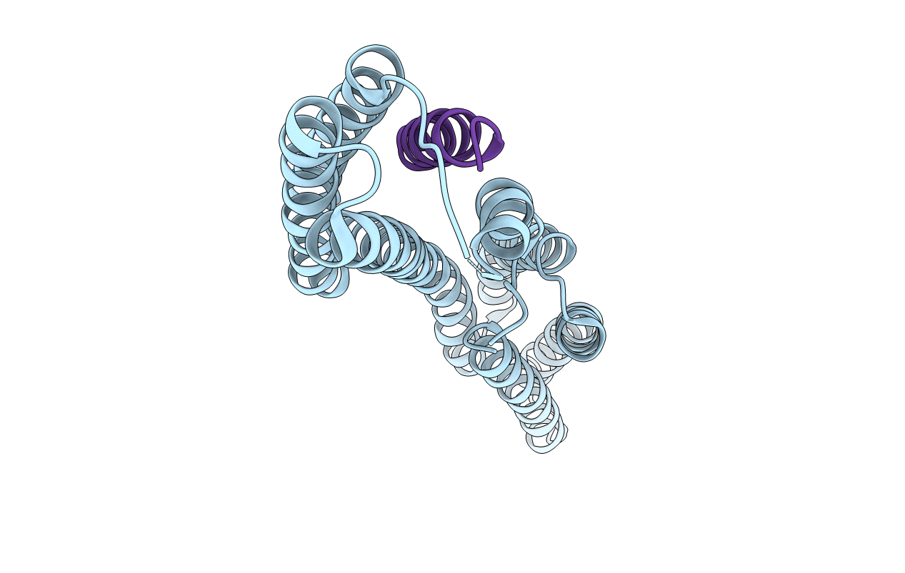
2GDC image
Deposition Date 2006-03-15
Release Date 2006-08-08
Last Version Date 2023-08-30
Entry Detail
PDB ID:
2GDC
Keywords:
Title:
Structure of Vinculin VD1 / IpaA560-633 complex
Biological Source:
Source Organism:
Gallus gallus (Taxon ID: 9031)
Shigella flexneri (Taxon ID: 623)
Host Organism:
Method Details:
Experimental Method:
Resolution:
2.74 Å
R-Value Free:
0.29
R-Value Work:
0.24
R-Value Observed:
0.24
Space Group:
P 21 21 2
Macromolecular Entities
Polymer Type:polypeptide(L)
Molecule:Vinculin
Gene (Uniprot):VCL
Chain IDs:A
Chain Length:266
Number of Molecules:1
Biological Source:Gallus gallus
Polymer Type:polypeptide(L)
Molecule:Invasin ipaA
Gene (Uniprot):ipaA
Chain IDs:B
Chain Length:21
Number of Molecules:1
Biological Source:Shigella flexneri
Primary Citation
Structural mimicry for vinculin activation by IpaA, a virulence factor of Shigella flexneri.
Embo Rep. 7 794 799 (2006)
PMID: 16826238 DOI: 10.1038/sj.embor.7400753

Abstact

Invasion of epithelial cells by Shigella flexneri is characterized by cytoskeletal rearrangements of the host cell membrane, promoting internalization of the bacterium. The bacterial effector IpaA is injected into the epithelial cell by a type III secretion apparatus and recruits vinculin to regulate actin polymerization at the site of entry. We analysed the complex formed between a carboxy-terminal fragment of IpaA (IpaA(560-633)) and the vinculin D1 domain (VD1), both in crystals and in solution. We present evidence that IpaA(560-633) has two alpha-helical vinculin-binding sites that simultaneously bind two VD1 molecules. The interaction of IpaA(560-633) with VD1 is highly similar to the interaction of the endogenous, eukaryotic proteins talin and alpha-actinin with VD1, showing that Shigella uses a structural mimicry strategy to activate vinculin.

Legend

Protein

Chemical

Disease

Primary Citation of related structures