2GD5 image
Entry Detail
PDB ID:
2GD5
Title:
Structural basis for budding by the ESCRTIII factor CHMP3
Biological Source:
Source Organism:
Host Organism:
PDB Version:
Deposition Date:
2006-03-15
Release Date:
2006-06-13
Method Details:
Experimental Method:
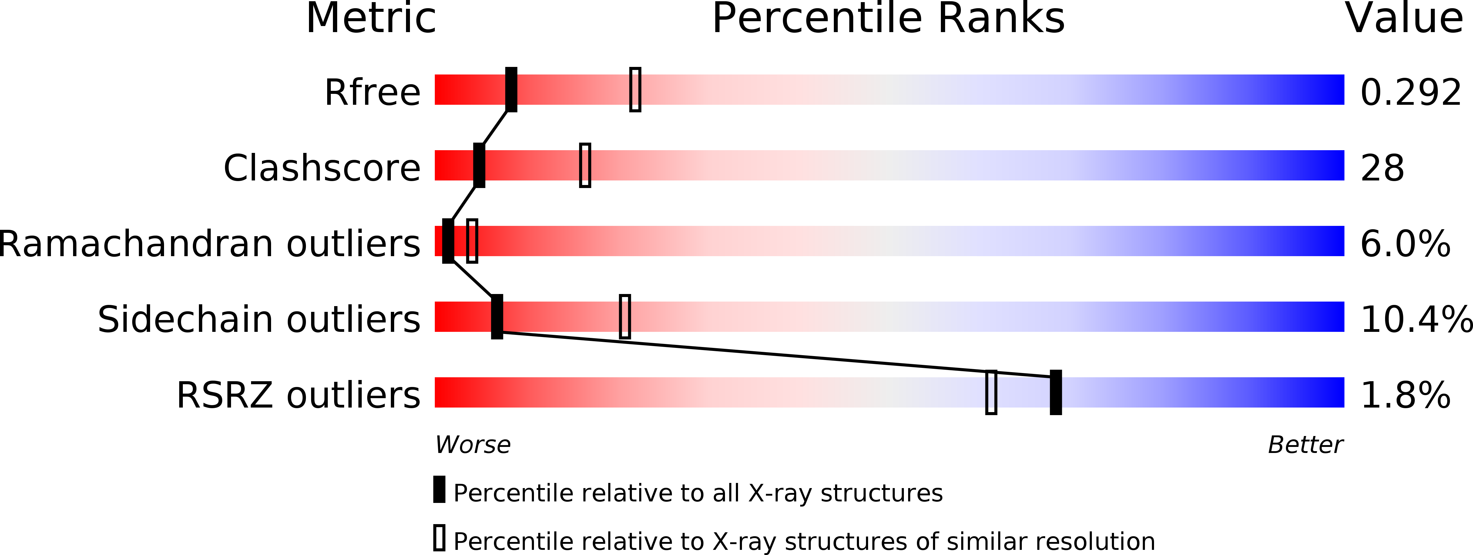
Resolution:
2.80 Å
R-Value Free:
0.30
R-Value Work:
0.25
R-Value Observed:
0.26
Space Group:
P 1
Macromolecular Entities
Polymer Type:polypeptide(L)
Description:Charged multivesicular body protein 3
Chain IDs:A, B, C, D
Chain Length:179
Number of Molecules:4
Biological Source:Homo sapiens
Modified Residue
Compound ID Chain ID Parent Comp ID Details 2D Image
MSE A MET SELENOMETHIONINE
Ligand Molecules
Primary Citation
Structural Basis for Budding by the ESCRT-III Factor CHMP3.
Dev.Cell 10 821 830 (2006)
PMID: 16740483 DOI: 10.1016/j.devcel.2006.03.013

Abstact

The vacuolar protein sorting machinery regulates multivesicular body biogenesis and is selectively recruited by enveloped viruses to support budding. Here we report the crystal structure of the human ESCRT-III protein CHMP3 at 2.8 A resolution. The core structure of CHMP3 folds into a flat helical arrangement that assembles into a lattice, mainly via two different dimerization modes, and unilaterally exposes a highly basic surface. The C terminus, the target for Vps4-induced ESCRT disassembly, extends from the opposite side of the membrane targeting region. Mutations within the basic and dimerization regions hinder bilayer interaction in vivo and reverse the dominant-negative effect of a truncated CHMP3 fusion protein on HIV-1 budding. Thus, the final steps in the budding process may include CHMP protein polymerization and lattice formation on membranes by employing different bilayer-recognizing surfaces, a function shared by all CHMP family members.

Legend

Protein

Chemical

Disease

Primary Citation of related structures